Some advice is best ignored. Like the warning Kyla Ortved received while volunteering in a small animal clinic as a teenager. One of the clinicians told her vet school was too hard to get into. The woman recommended Ortved do something else.
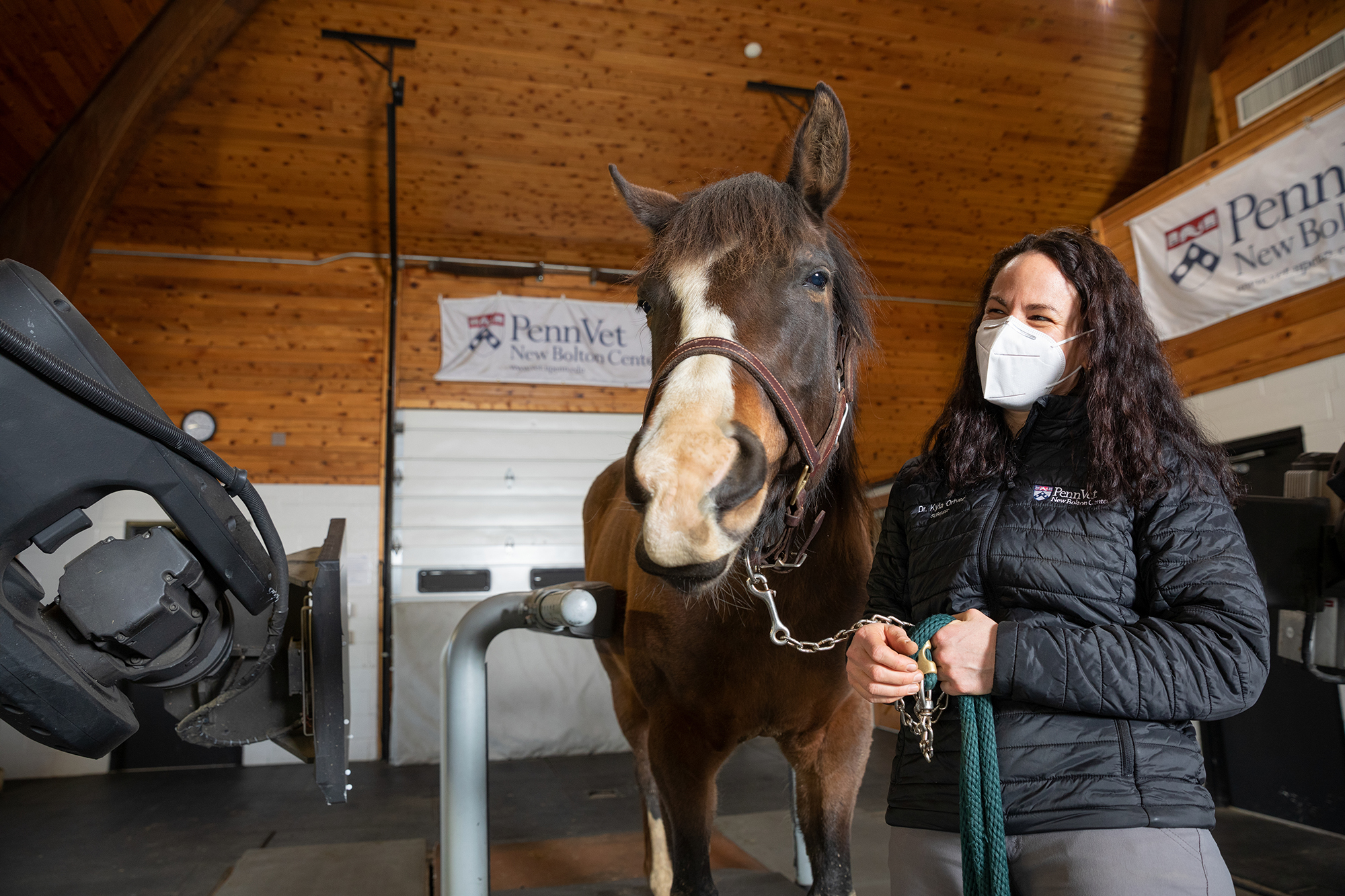
Fortunately for horse owners, students, and veterinary science in general, Ortved paid little attention. Twice board-certified—by the American College of Veterinary Surgeons and the American College of Veterinary Sports Medicine & Rehabilitation—today Ortved is a rising star in equine sports medicine and large animal surgery. And she runs the Ortved Laboratory at the School of Veterinary Medicine’s New Bolton Center, which studies post-traumatic osteoarthritis and explores regenerative medicine therapies to benefit horses and humans.
“I have a very specific memory of when I decided to specialize in surgery,” says Ortved, the Jacques Jenny Endowed Term Chair in Orthopedic Surgery. “During one of my externships, a resident invited me to participate in a procedure. The horse was under anesthesia for a check ligament desmotomy, a surgery to cut the check ligament for club feet. I helped with the surgery and realized: ‘This is it.’ And it still is. When I am doing surgery, I lose track of time. I’m where I’m meant to be.”
Up for a challenge
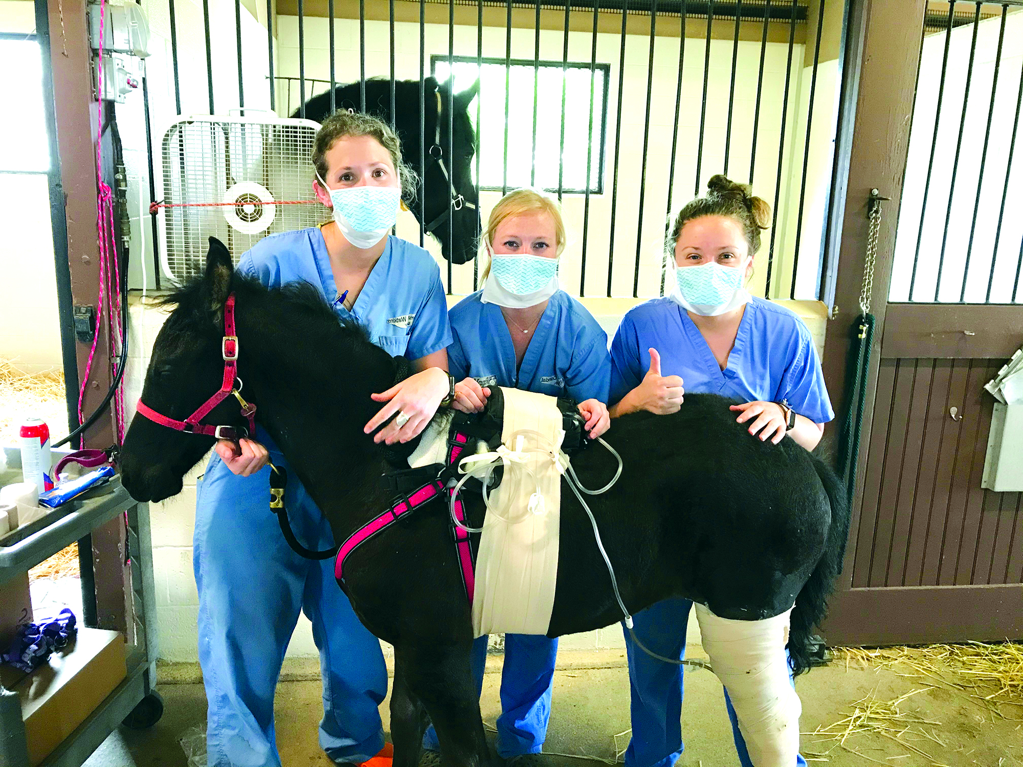
Recently, this place was with Osada, a two-month-old Friesian with a left tibial fracture.
“She was kicked by another horse,” says Daniel Lapp, owner of Red Crest Stables and breeders in Gordonville, Pennsylvania. “Our vet came to the farm to check her and asked if we’d want to pursue surgery, although she said it was a long shot.”
Lapp thought a long shot better than no shot, and the referring vet sent Osada’s X-rays to Ortved for an opinion.
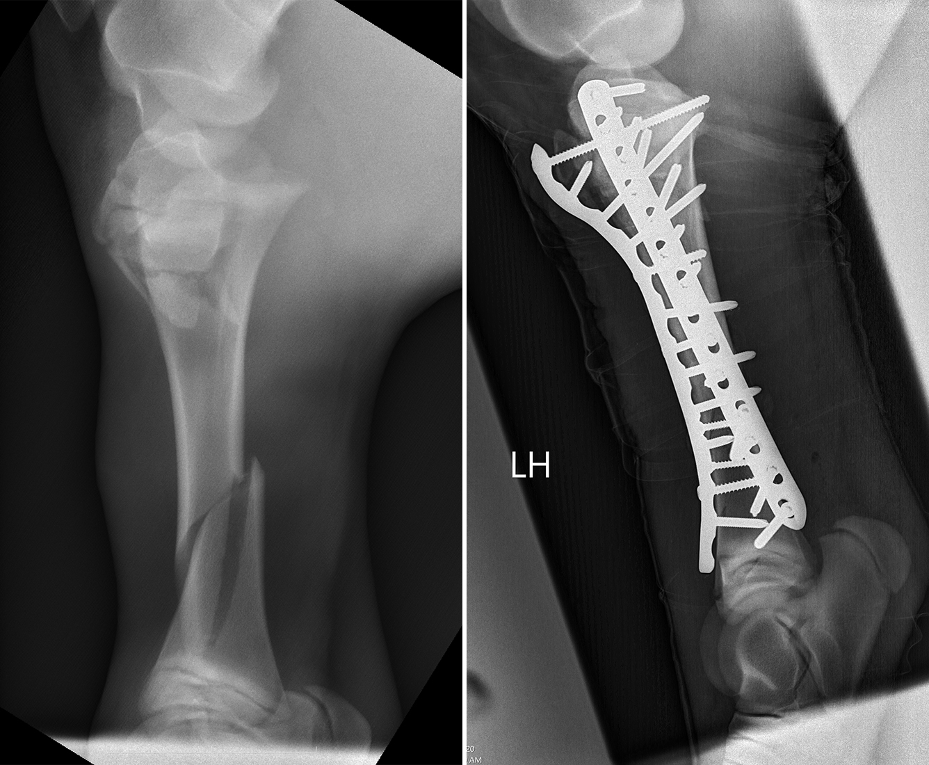
“It was a really severe fracture that was complicated by a second fracture at the growth plate at the top of the bone,” says Ortved. “In foals we usually see one or the other but rarely both. I knew it would be a bit more challenging to repair.”
She put Osada’s prognosis at 50%.
“I said go for it,” recalls Lapp.
Going for it
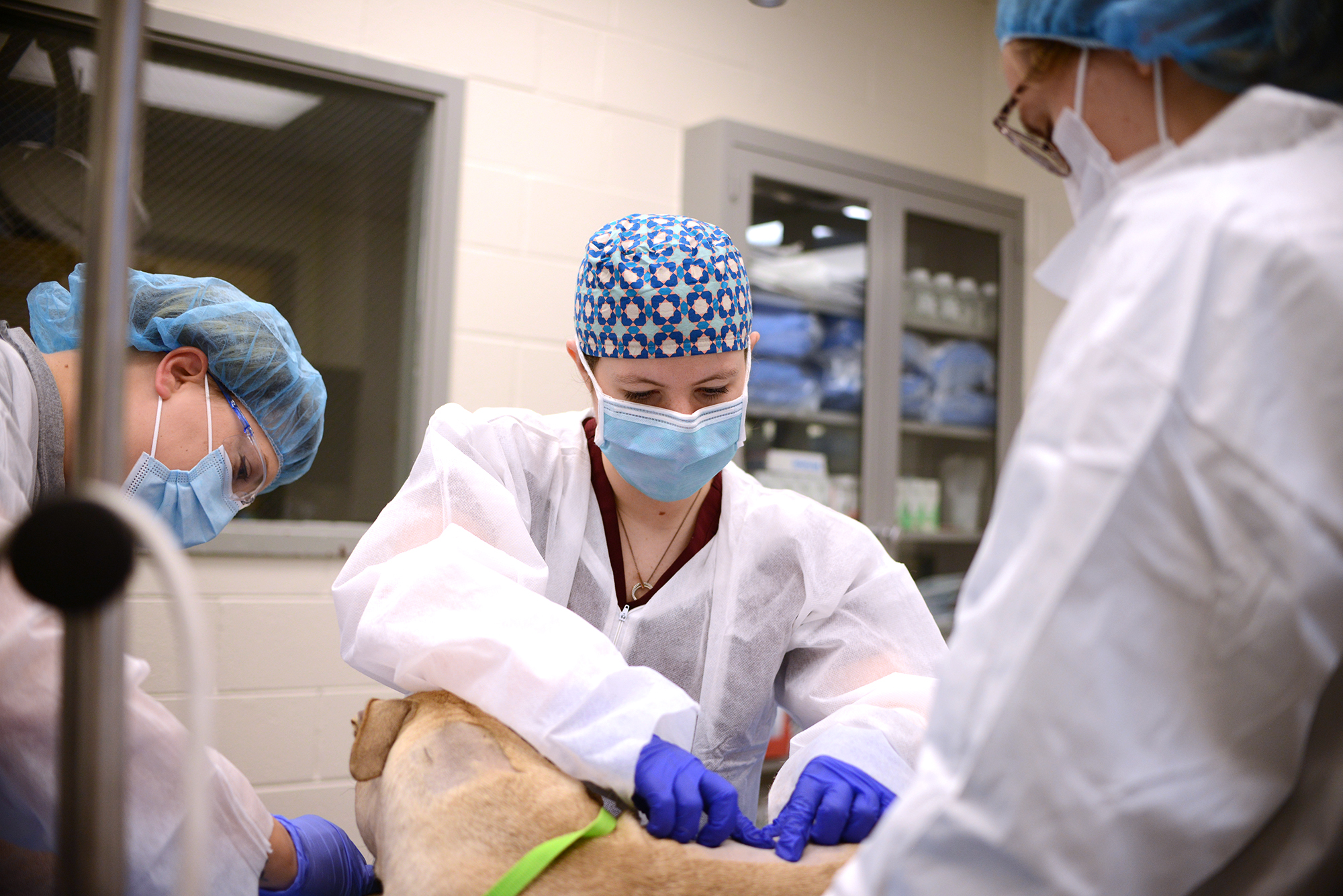
The New Bolton Center surgical team, which included radiology and anesthesia specialists and surgical nurses, prepped the horse. Dean Richardson, the Charles W. Raker Professor of Equine Surgery, also scrubbed in.
Osada was placed under general anesthesia. Two long, locking compression plates were carefully screwed into the bone in two places, a fracture repair approach for humans that Richardson adopted for equines early in his career.
After a lengthy, complex procedure—“the tibia is a hard bone to work with because access is difficult,” explains Ortved—they closed the incision in several layers, covering it with a light bandage.
The filly sailed through the surgery and subsequent recovery, but she wasn’t out of the woods. “The thing we’re most worried about in these cases is the implant becoming infected and the repair staying together,” Ortved says.
To mitigate the risk of infection, Osada received antibiotics, as well as anti-inflammatory medication and morphine for pain.
A few days post-op, Ortved’s fears came true. The wound opened and fluid leaked from Osada’s leg. The team cleaned the incision and performed a vacuum-assisted closure therapy to encourage drainage and healing. A bacterial culture found infection, so Ortved changed the horse’s antibiotics.
When Osada’s comfort didn’t improve, Ortved took her back to the operating room to lavage the wound. “She had an infection in the joint. We flushed the joint and placed antibiotic-impregnated bone cement in the wound to help with healing.”
The treatment worked. The wound started to close, and Osada slowly improved. She was discharged a few weeks after her last procedure.
21st-century surgery
“In the horse world, many people still think that fractures mean euthanasia,” Ortved says. “One of the takeaways from Osada’s case is that over the past several decades there have been so many advances in nursing care materials, in anesthesia, in surgical approaches that fractures don’t have to mean death. There are more options today.”
Cases like this one and legends like Richardson are among the top reasons Ortved joined New Bolton Center’s clinical team and faculty in 2016. A world-renowned surgeon, Richardson recently received the American College of Veterinary Surgeons ACVS Founders’ Award for Career Achievement for “contributions over the last 30+ years [that] have greatly impacted the art and science of veterinary surgery.”
Ortved says, “Working with Dean is an incredible opportunity and has vastly outweighed my expectation. I am unable to even come close to quantifying what I have learned at New Bolton Center and from him in the last five years.”
Raised on surgery
Ryan Hospital surgical resident Chiara Curcillo has conducted fewer surgeries than Ortved. But she has been around the specialty for many years, in a sense having grown up at Penn Vet.
Curcillo’s physician parents pioneered a single incision, minimally invasive laparoscopic surgical technique for humans. Jeffrey Runge brought it to animals when he was an assistant professor of surgery at Ryan Hospital.
“My parents would help Dr. Runge with the early procedures and take me along for the ride, knowing I wanted to be a veterinarian,” says Curcillo. “Dr. Runge guided me to where I am today. I spent several years shadowing him in the operating room [OR] and on the clinic floor. Having two surgeons raise me and a surgeon as my mentor made me want to be a surgeon and spend my days in the OR just like them.”
Quills, quills, and more quills
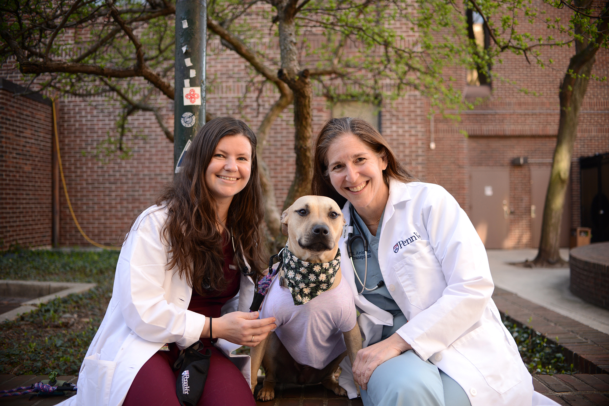
Curcillo had one of her trickiest surgeries yet with Daisy, an American Staffordshire Terrier who had an altercation with a porcupine right after Thanksgiving. The run-in resulted in what seemed like an endless number of quills stabbing the dog’s face and traveling throughout her body.
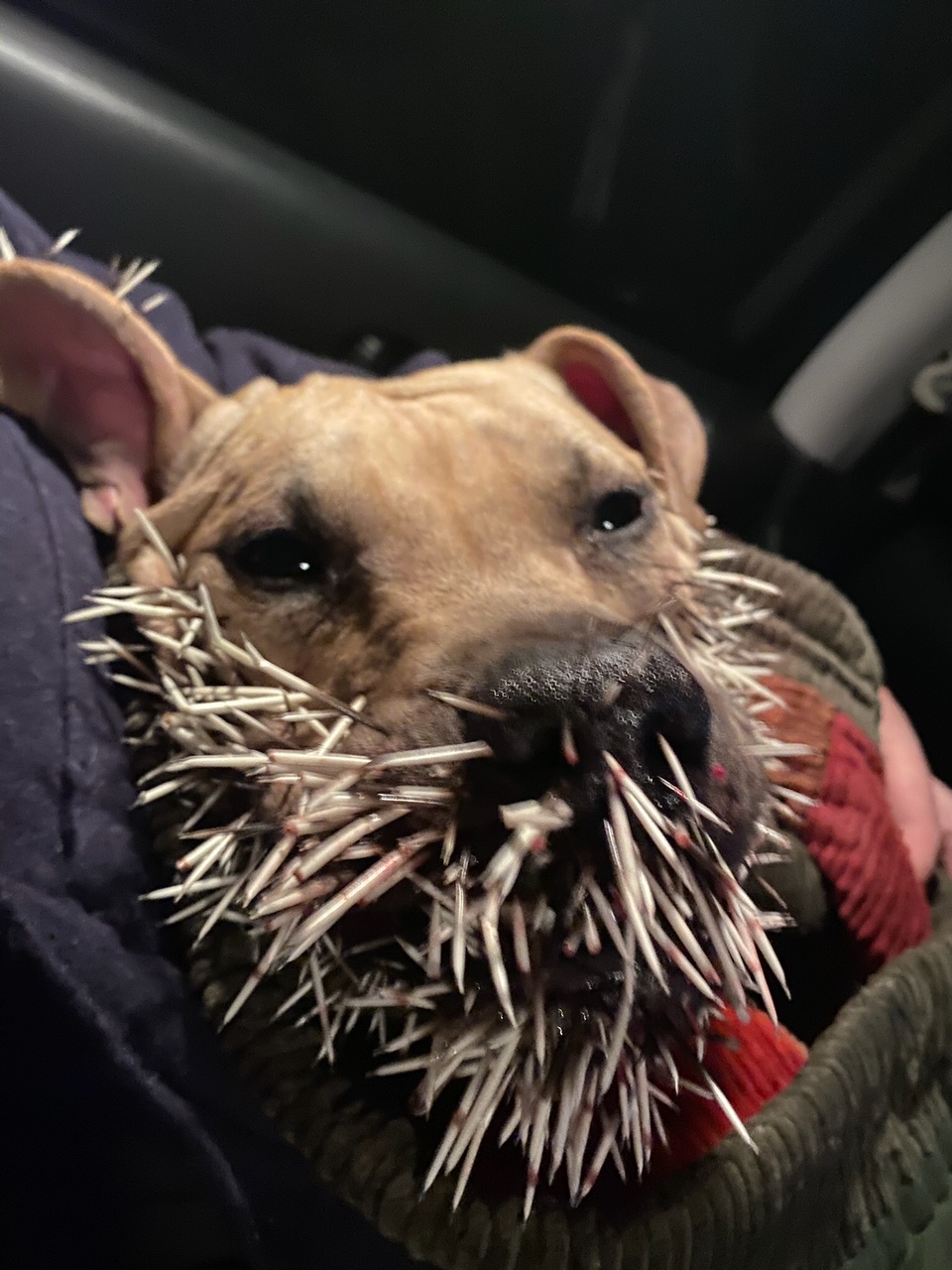
“We were visiting rural New York State and took her to an ER immediately, where they removed quills from all over her face and body,” says Katie Leonard, who, along with her husband, Franklin Donn, adopted Daisy in 2019. “As soon as we picked her up, we noticed more quills poking from her chin and pulled them.”
Additional quills surfaced when the couple, along with Daisy and Daisy’s canine brother Dexter, returned home to Baltimore.
But more worrisome symptoms overshadowed the quill appearances. Daisy’s breathing was labored, and she had lost interest in food and water.
At an ER in Baltimore, she was diagnosed with a pneumothorax, a dangerous condition of air in the chest cavity that can cause the lungs to collapse if untreated. Using a minimally invasive procedure called thoracocentesis, the ER vet placed a needle through Daisy’s chest wall into her chest cavity to remove air. Daisy was referred to a specialty practice, the third vet stop at this point.
Fourth and final stop
“Given the complexity of Daisy’s case, this last veterinarian then referred Daisy to us for further imaging and surgery,” Curcillo says.
This was the hope the family needed. “We weren’t feeling confident until we got to Ryan,” says Leonard. “Dr. Curcillo was very clear about the severity of Daisy’s situation but optimistic that all was not yet lost.”
After sedating the canine under general anesthesia and conducting a CT scan, Curcillo and Lillian Aronson, professor of surgery, opened Daisy’s chest and abdominal area. David Holt, professor of surgery, offered a hand.
Inside, the surgeons found a body riddled with quills—they were in the heart, lungs, liver, spleen, pancreas, kidney, diaphragm, and body wall.
“We removed each gingerly,” says Aronson. “We identified penetrating wounds in one of her lung lobes and performed a lung lobectomy. And we leak tested the other lobes, repairing a defect in one.” Before finishing the procedure, they placed a chest tube to help expel air.
Curcillo was working alongside the best: Aronson is a pioneer in small animal surgery and one of the first veterinarians in the world to perform renal transplants in dogs and cats—she’d conducted hundreds long before other surgical practices did any.
Also a noted small animal surgeon, Holt made news recently for charting an innovative approach to cancer surgery that helps surgeons clearly see whether they’ve left any diseased tissue behind in cancer excision surgery.
An ordeal continued
Post-op, Daisy woke up from anesthesia uneventfully and was transferred to the intensive care unit for monitoring. But, as with Osada, another hurdle was soon to come.
Day three after surgery, she developed abdominal discomfort and had fluid in her abdomen, as well as a suspected second pneumothorax.
It was back to surgery. Curcillo and Aronson opened her chest and abdomen again to evaluate the situation. After removing a few migrated quills, they examined her lungs, which had no apparent leaks, and replaced the old tube with a new one.
But air continued to escape post-surgery. In a minimally invasive procedure, “We then inserted an autologous blood patch to seal the sites in her lungs that were possibly leaking,” says Curcillo.
This did the trick. The rest of Daisy’s hospitalization progressed smoothly, and she was discharged a few days later.
A long and loving journey
“It’s been a long road for this sweet, outgoing dog,” says Curcillo. “But given her young age and that she is otherwise healthy, we believed this was a well-considered journey to take.”
Quills continue to surface even months after Daisy’s face-off with the spiky rodent—her owners estimate well more than 100 quills have been removed in total since November. And she has been back to Penn Vet several times for follow-ups and removal of aberrant spikes.
“Penn Vet is the only place we’ll go for our girl,” says Leonard. “We have wonderful pet insurance, thankfully. And Drs. Curcillo and Aronson and all of the clinicians who have worked so hard for Daisy are our ‘Team Daisy.’ We adore them.”
For Curcillo, Daisy is an unforgettable pup.
“I don’t know if I’ll ever see another case like this,” she says. “It was intense, challenging, and pretty cool—a perfect example of the type of surgery I’d hoped to experience at Penn Vet. Working next to Dr. Aronson and Dr. Holt is amazing—I am frequently in awe of them during a procedure. They teach me to be a great surgeon and a great educator. I aspire to be half the surgeons and teachers they are.”