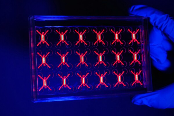
By applying tools of machine learning and network analysis, the Davis Lab in the Penn Epilepsy Center was assisted by a team of Penn interns this summer to target the ‘missing electrode problem,’ identifying regions of the brain that cause epilepsy.
One percent of the U.S. population, or three million people, lives with epilepsy. Approximately one-third of those will have a drug-resistant form of epilepsy that, often, demands surgery. The challenge: Localizing where their seizures are coming from before surgery has historically been done with the implantation of EEG electrodes in the brain—an extraordinarily invasive procedure that comes with its own set of limitations.
“The problem with intracranial EEG electrodes is mainly a sampling issue. To solve this problem, we are leveraging information captured from whole-brain neuroimaging to better localize the seizure onset zone,” says Andrew Revell, a fifth-year MD/Ph.D. student in the Davis Lab, who explains that implanted electrodes can miss the areas implicated in seizure generation. “When planning for epilepsy surgery to remove the seizure onset zone, you want to precisely localize the seizure generating areas and avoid eliminating any functional brain tissue, such as areas associated with hand movement or language generation. This is where an MRI can help.”
The Davis lab is studying how an MRI of the brain may help with the sampling issue of intracranial EEG, or the so-called “missing electrode problem,” where surgical limitations and safety restrict implantation of the entire brain. Patients routinely undergo an MRI of their brain before implantations, but they may also participate in research scans to acquire different imaging sequences, such as High-Angular Resolution Diffusion Imaging (HARDI). With these MRI sequences, the lab is modeling how the brain is connected, building networks of the brain, and making predictions of seizure activity in regions where electrodes were not implanted. They take an interdisciplinary approach, drawing from their expertise in imaging analysis, machine learning, network analysis, and signal analysis to solve a very relevant clinical problem.
Helpful in this process of innovating and improving the means of diagnosis for epilepsy is a team of Penn interns who, this summer, each took on different tasks related to this larger project. Revell describes their work as “tremendously helpful and a very rewarding experience for the lab,” addressing the need for a team of scientists and students to take on an important clinically translational task.
“They bring in new perspective and challenge me as a Ph.D. student,” he says. “I have grown a lot over the summer in mentoring them, and I hope they have learned and taken away from the program just as much as I have.”
“The goal of this project is to predict the function of the brain measured with the intracranial EEG implantations with structural neuroimaging of the brain [HARDI].” Many patients continue having seizures after epilepsy surgery and this project could answer the question of, ‘What went wrong?’” Armstrong says. “This could be that next frontier of options. We can have a better understanding of seizures if we better understand the brain’s structure-function relationship.”
Dhanya Mahesh, a senior sponsored by MindCORE, examined the differences between white matter and grey matter electrode recordings. Newer implantation methods adopted at Penn record neural activity in both gray matter and white matter tissue. It is currently unknown how measurements recorded in both tissue types may affect current models of the brain’s structure-function relationship. Furthermore, it is currently unknown if epileptologists should treat grey matter and white matter signals differently when interpreting EEGs.
“My project compared those two readings using the signal’s perspective to figure out if maybe the grey area is giving more seizure-related info, or giving the same info as the white area,” Mahesh says. “Supposing they are giving the same info, we may only need to consider gray matter electrodes.”
She describes it as a less-studied area and says it could have real implications for how surgeons and epileptologists determine where a seizure is beginning and ending. She was thrilled to have the opportunity, she says, especially because she chose Penn because of its relationship with Penn Medicine.
Kevin Matthews, meanwhile, is also a sophomore who joined the lab through CURF and was part of PURM; he was mentored by Campbell Arnold, another Ph.D. student in the Davis lab. A dual major in behavioral economics and computer science, Matthews examined the relationship between brain age and epilepsy patients. The idea was that brain age could be a useful determinant for who is a good candidate for surgery. However, preliminary results from his summer project showed that brain age is not a statistically significant indicator, which conflicts with prior research on brain age in other neurological disease studies. This work is ongoing.
In the process, Matthews says he learned a lot about how servers and different machine learning algorithms work./p>
“I was interested in the machine learning of this and the statistical aspects,” he says of his internship experience. “I would never have guessed I’d be applying my computer science skills in a medical context,” he adds, “but it’s really interesting and I’m considering some other research that’s similar.”
Ellie Chen, a junior electrical engineering and junior computer science major from Dallas, was determined to apply her interest in engineering to neuroscience and find a lab where she could do so. Her work in the lab, sponsored by the Littlejohn Undergraduate Research Program, looks at networks in the brain and where seizures travel. That research has since evolved to an ongoing new project examining lower-field 3T MRI scanners that could potentially be used as a substitute for 7T MRI scanners in either screening or diagnostics, taking lower-quality scans and using machine learning to fill in the gaps. It would have big implications for accessibility if the lower-field scans are found to be useful, she says.
“I’ve been really interested in research and it’s cool to be involved in a project and given responsibility as an undergrad,” Chen says. “You get to see the impact of your project.”
Davis says she was blown away by the contributions of the undergraduate students over the summer, particularly in light of the need for a virtual lab experience during the COVID-19 pandemic. She credits the overwhelming success of the students to her graduate students and each of the students’ dedication, enthusiasm, and aptitude.
“The tremendous progress that was made over the summer in our work is a testament to the outstanding Penn student body and the multitude of summer intern programs for undergraduates,” she says. “The Penn campus is uniquely integrated with the medical campus, which accelerates translational research. The majority of the summer students have chosen to stay on in my lab during the school year, which is ideal. I’m very much looking forward to mentoring them throughout the year and beyond.”
Penn engineers and collaborators have developed a transparent, micro-engineered device that houses a living, vascularized model of human lung cancer—a “tumor on a chip”—and show that the diabetes drug vildagliptin helps more CAR T cells break through the tumor’s defenses and attack it effectively.
Tumor-on-a-chip offers insight into cancer-fighting cells in immunotherapy
Penn engineers and collaborators have built a living tumor on a chip to expose how cancers block immune attacks, and how one existing drug could make immunotherapy like CAR T more effective against solid tumors.
Professor of city and regional planning Erick Guerra recently published a book exploring the economic and societal impacts of American highways. He explains some of the pitfalls associated with an ever-expansive highway system, arguing that spending more on highways might not be the solution to the country’s transportation issues.
Penn urban planner Erick Guerra’s new book, “Overbuilt,” argues that additional spending on building more highways might not be the solution to the country’s transportation issues. In a Q&A, Guerra shares his insights.
Xin Sun prepares samples collected from the Eastern Tropical North Pacific aboard a research vessel. By adding stable isotope tracers to these vials, Sun and her team can track how different microbial groups convert nitrogen compounds into nitrous oxide, revealing how subtle shifts in oxygen and organic matter change the ocean’s chemistry.
Can tiny ocean organisms offer the key to better climate modeling?
In the shadowy layers of the Pacific, microbes decide how much nitrous oxide—a potent greenhouse gas—rises skyward. New research from Penn’s Xin Sun offers an improved understanding of microbial ecology and geochemistry—key to forecasting global emissions in response to natural and man-made climate change.
Two X-ray plates from Arthur Goodspeed, believed to have created the world’s first X-ray image, were donated by his family to Penn’s University Archives.


