New Multi-photon Microscope at Penn Vet Offers Faster, Deeper Images
By Patrick Ammerman
You can’t study what you can’t see.
That is why the University of Pennsylvania School of Veterinary Medicine’s Imaging Core is investing in a multi-photon microscope. The new device, which specializes in capturing 3-D images of thick living and fixed tissues, is to be installed at Penn Vet this summer and will enhance the bio-imaging infrastructure there and in Penn’s Perelman School of Medicine.
[vimeo]175688770[/vimeo]
The microscope will further many core areas of research in Penn Vet and across the University.
The microscope uses advanced lasers to excite labeled molecules in a biological sample. It can simultaneously detect as many as four different signals in a scan. For instance, the instrument could capture the interaction between a parasite labeled with a green fluorescent molecule and a lymphocyte of the immune system labeled with red. At the same time, it could track the cell with which the lymphocyte was interacting, marked with yellow, and keep tabs on intracellular calcium, a signaling molecule, with a blue reporter.
The microscope’s computer can rapidly reconstruct 3-D images of these signals, allowing researchers to view cells or molecules of interest as they move and respond in living tissue. Observing all of these facets interacting in real time enables researchers to obtain a clearer understanding of the mechanisms that underlie important biological functions and the pathogenesis of disease.
According to Bruce Freedman, an associate professor in Penn Vet’s Department of Pathobiology and director of the Imaging Core facility, multi-photon microscopy is crucial to the work of many researchers in labs across Penn, including, in addition to Penn Vet and Perelman, the School of Dental Medicine and the School of Arts & Sciences.
Due to its imaging abilities, this new instrument “will have transformative impact on our understanding of biology in vivo,” Freedman said.
The new microscope replaces an instrument that the Core acquired in 2008. This older 2-photon instrument is currently used for research projects in immunology, host-microbial interactions, germ cells, musculoskeletal systems, oncology and ophthalmology. More than 80 peer-reviewed publications have been attributed to the Penn Vet 2-photon microscope.
Among its critical advantages is its ability to image more deeply into specimens and to acquire images more rapidly with less noise and by inflicting less damage on the tissue sample. Freedman estimates that it can scan four to six times deeper into tissue than the older unit.
Freedman said the new scope will better equip Penn researchers who are pushing against the technical and intellectual frontiers of science.
“Maybe there was some event that we would miss with this old microscope, and now it’s within the capability of the new one to capture that event,” he said. “We can now visualize cell behaviors as they occur in real time with much greater resolution in three dimensions and over longer times.“
Freedman pointed to the microscope’s ability to capture events taking place inside organs like the lymph node or the brain as applications of its greater imaging power.
Freedman uses 2-photon microscopy as a part of his own research on cellular and molecular interactions in the thymus, a gland that that is part of the immune system that helps prevent autoimmunity.
While cells in the thymus can be viewed in vitro, Freedman said that 2-photon microscopy is crucial to understanding how the gland actually instructs the immune system so that it can recognize pathogens in living organisms.
“We all use study cell models of disease in dishes and incubators, but ultimately what we really want to do is this: we want to just see what happens in real life, in vivo, in an unperturbed environment.”
The microscope purchase is being funded in large part by a grant from the National Institutes of Health, with additional support from Penn Vet.
In Senegal, the ambitious Dakar Greenbelt project seeks to create an extensive network of ecological infrastructure in and around the city to sustainably address environmental concerns and enhance urban life. With support from David Gouverneur and Ellen Neises, Ph.D. candidate Rob Levinthal in the Weitzman School of Design led two courses that included a field trip to Dakar, that culminated in students presenting their visions for parts of the Greenbelt.
From a desert to an oasis: Penn engages in ambitious greening effort in the Sahel
Students from the Weitzman School of Design journeyed to Senegal to help with a massive ecological and infrastructural greening effort as part of their coursework. The Dakar Greenbelt aims to combat desertification and promote sustainable urban growth.
As part of an undergraduate course, Penn faculty and students curated an Arthur Ross Gallery exhibition of works from the Neumann family’s extensive collection of modern and contemporary art.
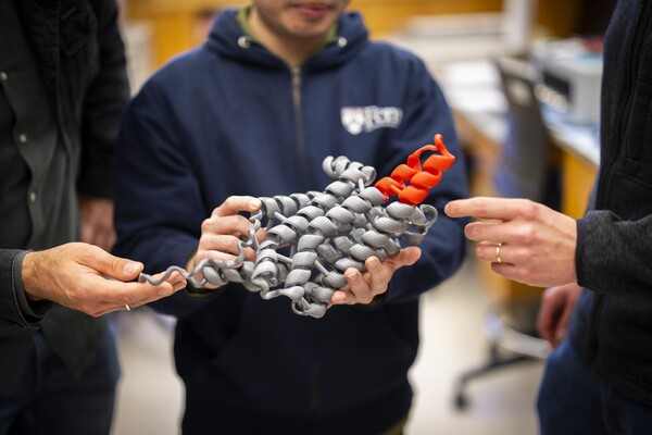
The University’s nexus for technology transfer supports researchers in their innovative efforts, from CAR T to mRNA advancements that have dramatically reshaped the world.