(From left) Doctoral student Hannah Yamagata, research assistant professor Kushol Gupta, and postdoctoral fellow Marshall Padilla holding 3D-printed models of nanoparticles.
(Image: Bella Ciervo)
Shuying (Sheri) Yang, a new associate professor in the University of Pennsylvania School of Dental Medicine’s Department of Anatomy & Cell Biology, began her career as a medical student, the fulfillment of a childhood dream. But during her five years of medical school, she realized the many limitations and challenges to medical treatment for the different diseases she was encountering in the clinic and hoped to help advance treatment options. So she made the decision to continue her education, pursuing graduate degrees, in basic research on gene therapy, cancer biology and, eventually, bone biology.
That foray into research shaped her career, which she has spent largely in dental schools, contributing to an understanding of the molecular mechanisms of bone development, remodeling and repair. She’s also used her insights to identify therapeutic targets to treat bone diseases such as osteoporosis and fracture.
“Dr. Yang brings a strong portfolio of research and scholarship to the School and her work in bone development has potential for impacting patient care not only within the field of dental medicine but beyond,” says Denis Kinane, Penn Dental Medicine’s Morton Amsterdam Dean, on her appointment this June. “We are pleased to have her here at Penn Dental.”
Yang pursued a postdoctoral fellowship in the lab of Renny Franceschi in 2000 at the University of Michigan, where she started her bone biology study. There, together with colleagues, she provided the first evidence that host cells are major contributors to bone regeneration and repair following stem cell and BMP2 gene modified fibroblast cell transplantation into the areas where a defect existed. They discovered that Runx2 and BMP2 have distinct, but complementary, roles in controlling both the extent and type of bone formed in regenerating sites and that their combined action is necessary for optimal bone formation.
To expand her focus and obtain additional training in molecular and genetic techniques, she joined Yi-Ping Li’s lab at the Forsyth Institute and Harvard School of Dental Medicine in 2002, where she remained for six and a half years. Yang’s work resulted in the publication of a number of seminal papers on the cloning and characterization of genes critical to osteoclast differentiation and function. In particular, her research revealed for the first time that RGS10 and RGS12, regulators of G protein signaling, were critical regulators for osteoclast differentiation. The findings from that time earned her awards in the bone field, including the American Society for Bone and Mineral Research’s Most Outstanding Abstract Award and the ASBMR John Haddad Young Investigator Award in Advances in Mineral Metabolism.
“That was a great period of time for me, enabling me to develop skills, including study design and management, grant writing, and to master molecular genetics, genomics and cell biology knowledge and techniques,” Yang says. “Those experiences readied me to become an independent investigator.”
The opportunity to run her own lab came at the State University of New York at Buffalo School of Dental Medicine, which she joined in 2008.
As she set up her own lab, Yang focused in on three main research thrusts, each with a basic and a clinical science component, true to her early interest in translational medicine.
First, she and her lab have examined the role of regulators of G protein signaling in bone development, bone remodeling and aging. While ample research has shown that these proteins play critical roles in normal physiology as well as in pathological conditions in immune cells, neurons and cardiac myocytes, among others, their role in bone cells has remained unexplored.
“In the bone field, how RGS proteins regulate bone remodeling and what role they play in the aging skeleton is largely unknown,” says Yang.
Using a genome-wide screening method, Yang and colleagues identified the largest protein in the regulators of G signaling proteins family, RGS12, which plays a pivotal role in osteoclast differentiation. They then developed conditional knockout mouse models to define the role of RGS12, finding it to be essential for bone resorption and formation. Yang discovered that deletion of RGS12 in osteoclast cells caused mice to have greater bone mass and less age-related inflammatory bone loss. She is hopeful that the work, supported by the National Institutes of Health, will lead to new ways to target RGS12 or other RGS12-interacting proteins involved in bone remodeling as a treatment for conditions that involve bone diseases, such as age-related osteoporosis and osteoarthritis.
A second major emphasis in Yang’s research has been a focus on intraflagellar transport , or IFT, proteins, which are essential for the creation of the cilia, microtubule-based sensory organelle that extends from the cell body.
“Back when I was in medical school, I learned that each cell has only one primary cilia, but we didn’t know about their function,” Yang says. “From one of the publications of my current collaborator, Rosa Serra, I was excited to find that defective ciliary-related proteins can cause severe craniofacial and bone disorders. That was the first time I realized, wow, this small, hair-like structure is so important in bone. I became very interested in this study.”
Indeed, though cilia were first discovered in chondrocytes in the 1960s, subsequent work has revealed that cilia are also present in osteoblasts, osteocytes and mesenchymal stem cells. It wasn’t until the 2000s that the study of ciliopathies, disorders that cause a wide range of syndromic maladies that involve nearly every major body organ, including the kidney, brain, limb, retina, liver and bone, and cilia’s role in bone disorders became more apparent.
“Although the involvement of primary cilia and IFT proteins in human diseases is now well established, there are still many unanswered questions about their function and biological mechanisms,” Yang says.
Using molecular, cellular and genetic approaches, she discovered that the IFT protein, which constructs cilia by shuttling proteins back and forth to the growing cilium’s tip, also helped regulate bone development and bone remodeling through balancing hedgehog canonical and non-canonical signaling pathways, a finding recently published in Nature Communications.
One intriguing line of research in this area involves looking at the cilia as a mechanical sensor.
“We know if we are sedentary, we get fatter and experience bone degeneration or loss, and, if we do exercise, our bones become stronger,” Yang says. “But we don’t understand exactly how that happens. We would like to know how important cilia and IFT proteins are for mechanical signaling in bone and whether and how IFT protein are involved in mechanical signal transduction to drive stem cells to become osteoblasts instead of adipocyte to promote bone formation. This study will lead us to find new insight to prevent losing bone and gaining fat.”
A final set of studies in Yang’s lab has a more direct applied aim: to develop new types of grafts to repair large bone defects. She notes that while small bone defects and injuries can be quickly healed, often without much clinical intervention, large ones, such as those that arise following a large trauma or that remain after a tumor is removed, have remained stubbornly difficult to fix, with clinicians resorting to artificial materials to bridge defects.
The problem is not just that bone cells fail to regenerate enough to bridge the gap but the absence of blood vessels.
“If there is no blood, there is no nutrition and bones cannot be regenerated,” Yang says.
Through a collaboration with researchers at SUNY-Buffalo, she is experimenting with different growth factors, stem cells and biodegradable and injectable scaffolds that promote both angiogenesis and osteogenesis. The challenge has been to release growth factors slowly, in a long-term fashion to allow the bone to form gradually.
“So we’re trying to think about how we can optimize the conditions, using a growth factor-infused scaffold so the growth factor is slowly released and can stay in the body for a longer time, actively stimulating bone regeneration,” says Yang.
Since arriving at Penn Dental Medicine, Yang has been exploring collaborations with her colleagues within the School, notably Songtao Shi, professor and chair of the Department of Anatomy & Cell Biology, who has also investigated the mechanisms and applications of stem cell therapy and differentiation, and Dana Graves, professor and interim chair of the Department of Periodontics, who has examined the molecular basis of bone remodeling and in particular how diabetes affects bone.
She’s also looking forward to taking advantage of the core facilities and other researchers’ expertise from around the University to advance her work on how bone cells use their cilia to process mechanical signals, including Robert Mauck of the Perelman School of Medicine, who directs the Biomechanics Core of the Penn Center for Musculoskeletal Disorders.
“Many dentists work in the clinic, but at the dental school and all around Penn you find people who are also deeply involved in research,” Yang says. “I feel it’s a very unique environment in which to do research.”
Katherine Unger Baillie

(From left) Doctoral student Hannah Yamagata, research assistant professor Kushol Gupta, and postdoctoral fellow Marshall Padilla holding 3D-printed models of nanoparticles.
(Image: Bella Ciervo)

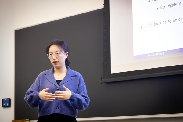
Jin Liu, Penn’s newest economics faculty member, specializes in international trade.
nocred

nocred

nocred