Penn Researchers Develop Biological Circuit Components, New Microscope Technique for Measuring Them

PHILADELPHIA — Electrical engineers have long been toying with the idea of designing biological molecules that can be directly integrated into electronic circuits. University of Pennsylvania researchers have developed a way to form these structures so they can operate in open-air environments, and, more important, have developed a new microscope technique that can measure the electrical properties of these and similar devices.

The research was conducted by Dawn Bonnell, Trustee Chair Professor and director of the Nano/Bio Interface Center, graduate students Kendra Kathan-Galipeau and Maxim Nikiforov and postdoctoral fellow Sanjini Nanayakkara, all of the Department of Materials Science and Engineering in Penn’s School of Engineering and Applied Science. They collaborated with assistant professor Bohdana Discher of the Department of Biophysics and Biochemistry at Penn’s Perelman School of Medicine and Paul A. O’Brien, a graduate student in Penn's Biotechnology Masters Program.

Their work was published in the journal ACS Nano.

The development involves artificial proteins, bundles of peptide helices with a photoactive molecule inside. These proteins are arranged on electrodes, which are common feature of circuits that transmit electrical charges between metallic and non-metallic elements. When light is shined on the proteins, they convert photons into electrons and pass them to the electrode.

“It's a similar mechanism to what happens when plants absorb light, except in that case the electron is used for some chemistry that creates energy for the plant,” Bonnell said. “In this case, we want to use the electron in electrical circuits.” 

Similar peptide assemblies had been studied in solution before by several groups and had been tested to show that they indeed react to light. But there was no way to quantify their ambient electrical properties, particularly capacitance, the amount of electrical charge the assembly holds.

“It’s necessary to understand these kinds of properties in the molecules in order to make devices out of them. We've been studying silicon for 40 years, so we know what happens to electrons there,” Bonnell said.  “We didn’t know what happens to electrons on dry electrodes with these proteins; we didn't even know if they would remain photoactive when attached to an electrode.” 

Designing circuits and devices with silicon is inherently easier than with proteins. The electrical properties of a large chunk of a single element can be measured and then scaled down, but complex molecules like these proteins cannot be scaled up. Diagnostic systems that could measure their properties with nanometer sensitivity simply did not exist.

The researchers therefore needed to invent both a new way of a measuring these properties and a controlled way of making the photovoltaic proteins that would resemble how they might eventually be incorporated into devices in open-air, everyday environments, rather than swimming in a chemical solution.  

To solve the first problem, the team developed a new kind of atomic force microscope technique, known as torsional resonance nanoimpedance microscopy. Atomic force microscopes operate by bringing an extremely narrow silicon tip very close to a surface and measuring how the tip reacts, providing a spatial sensitivity of a few nanometers down to individual atoms.  

“What we've done in our version is to use a metallic tip and put an oscillating electric field on it. By seeing how electrons react to the field, we’re able to measure more complex interactions and more complex properties, such as capacitance,” Bonnell said.

Bohdana Discher’s group designed the self-assembling proteins much as they had done before but took the additional step of stamping them onto sheets of graphite electrodes.  This manufacturing principle and the ability to measure the resulting devices could have a variety of applications.  

“Photovoltaics — solar cells — are perhaps the easiest to imagine, but where this work is going in the shorter term is biochemical sensors,” Bonnell said. 

Instead of reacting to photons, proteins could be designed to produce a charge when in the presence of a certain toxins, either changing color or acting as a circuit element in a human-scale gadget.  

This research was supported by the Nano/Bio Interface Center and the National Science Foundation.


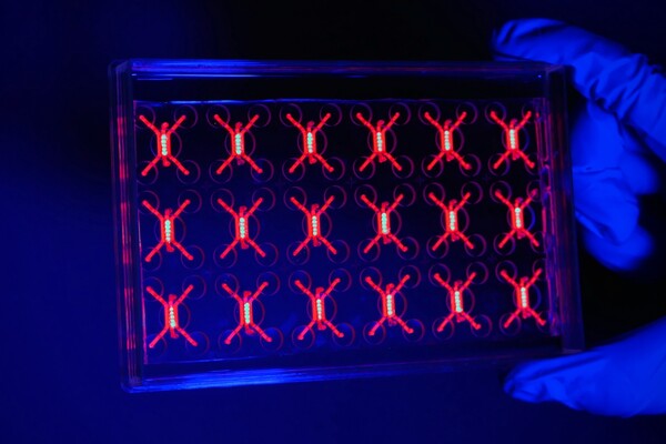
Image (above right) reprinted with permission from "Direct Probe of Molecular Polarization in De Novo Protein–Electrode Interfaces," Kendra Kathan-Galipeau, Sanjini Nanayakkara, Paul A. O’Brian, Maxim Nikiforov, Bohdana M. Discher, Dawn A. Bonnell, ACS Nano, Copyright 2011 American Chemical Society