Penn Vet Study Identifies Mechanism Explaining Female Bias in Autoimmunity
Possessing two X chromosomes is a double-edged sword, immunologically speaking. Females are better at fighting off infection than males, but they are also more susceptible to many autoimmune conditions, such as lupus.
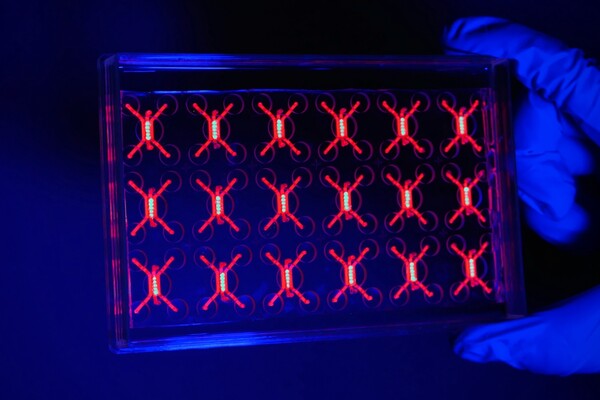
The research team, headed by Montserrat C. Anguera, an assistant professor in the Department of Biomedical Sciences at Penn’s School of Veterinary Medicine, focused on X inactivation, a mechanism that serves to balance gene dosage between males and females by inhibiting gene expression on one of a female’s two X chromosomes. They discovered that, in females, the immune system’s lymphocytes, specifically T cells and B cells, lack the typical patterns of Xist, an RNA molecule essential for the inactivation process, as well as other markers of X chromosome inactivation, thus making X inactivation incomplete in these cells.
That incomplete activation was present in lymphocytes in all females, but lupus patients also had unique expression patterns of key immunity-related genes and unusual patterns of Xist RNA localization, suggesting an underlying explanation for the disease condition.
“There just seems to be something about lymphocytes,” Anguera said. “The silencing of the X chromosome doesn’t seem to be as tight in them as it is in other cell types.”
Anguera’s coauthors on the paper were lead author Jianle Wang, Camille Syrett and Michael Atchison, all of Penn Vet; Marianne C. Kramer of the Department of Biochemistry and Biophysics in Penn’s Perelman School of Medicine; and Arindam Basu of Pennsylvania State University, formerly of Penn Vet.
The study had its origins in Anguera’s postdoctoral research, which focused on X inactivation in pluripotent stem cells. In particular she examined the role of Xist, a long non-coding RNA molecule that is known to initiate X inactivation and maintain it by residing with the inactivated X chromosome, which also acquires small-molecule tags called heterochromatic modifications, further repressing gene expression.
Anguera’s studies found that, in some cases, stem cells lose both Xist expression and the modifications that normally reside on the inactive X. As a result, the cells would start to become partially reactivated, growing rapidly and beginning to resemble cancer cells.
She began to wonder whether certain diseases might arise from improper maintenance of X inactivation, notably autoimmune conditions such as lupus; 85 percent of lupus patients are women.
“What caught my attention about autoimmunity and specifically lupus,” Anguera said, “was that there were genes on the X chromosome that were immunity related and that had been shown to have higher expression levels in lupus patients.”
To address this question, the Penn researchers examined lymphocytes donated by healthy human females as well as “naïve,” or unstimulated T and B cells from female mice. They found that, unlike in other cell types where Xist is associated tightly with the inactive X chromosome, female lymphocytes lacked this Xist “cloud,” even though the cell still contained expected levels of Xist. This suggested that Xist was failing to properly migrate to the inactive X to silence it.
When the research team activated the human T and B cells, simulating how these cells would respond when presented with a pathogen, the Xist clouds reappeared. The team observed similar results in mice. Yet the naïve and stimulated lymphocytes all had similar quantities of Xist.
“What was really striking to us was that this wasn’t due to a difference in the amount of Xist. There is a ton of Xist RNA in these cells,” Anguera said. “It’s just not getting to the inactive X chromosome in the naïve lymphocytes.”
Further examinations of the inactive X in lymphocytes found that it lacked the heterochromatic marks found on the inactive X in other cell types.
The findings that even healthy females had such unusual maintenance of the inactive X in their lymphocytes was entirely unexpected.
“Our hypothesis was that the lupus samples were going to be dysregulated and the healthy females would be fine,” Montserrat said. “So it was really shocking to us that lymphocytes in normal females lacked these markers of X inactivation as well.”
To see if this lack of heterochromatic marks and Xist, both of which normally block gene expression on the inactive X, resulted in increased gene expression, the researchers looked at immunity-related genes on the X chromosome to see if there was one or two copies being expressed. They found that about 3 to 5 percent of female lymphocytes expressed two copies of these genes, as well as two copies of a gene unrelated to immunity. No male lymphocytes showed this same two-copy expression.
Consistent with that result, the researchers found that certain regions of the X chromosome in human female B cells, including regions that contain immunity-related genes, were expressed at higher levels than male cells.
Because Xist in the female lymphocytes was present but simply wasn’t localizing to the proper place on the inactive X, the research team took a closer look at two proteins, YY1 and hnRNPU, that are known to bind with Xist and possibly play a role in moving it back to the inactive X after lymphocytes are stimulated. Using human T cells in culture as well as mice lacking one of these genes, YY1, they found that, indeed, the two proteins did help move Xist back to the inactive X chromosome in activated lymphocytes.
Though it was clear that female lymphocytes were different from males’ in their patterns of Xist localization, the team wanted to know whether lupus patients had additional unusual features of X inactivation that might explain their disease. Using lymphocyte samples from both pediatric lupus patients and healthy children of similar ages, the team found more Xist mis-localization in lupus patients and some evidence that they were more likely than healthy people to have two copies of immunity-related genes. The researchers also noted that the region of the X chromosome containing Xist showed the biggest differences in expression when comparing lupus to healthy patients.
One possibility, said Anguera, is that all females may have a subpopulation of lymphocytes with incomplete X inactivation; in healthy individuals, those lymphocytes stay in the minority, but that subpopulation may take over in patients with autoimmune conditions.
To build on the work, Anguera’s team is performing additional studies using primary samples from lupus patients as well as samples from a mouse model of lupus. While the current study was done on immortalized cells from lupus patients, Anguera is hopeful the researchers will obtain a clearer picture of how Xist patterns differ using primary cells.
The team is also embarking on a study of another female-biased autoimmune disease, Sjogrens syndrome, which has an even more extreme female bias than lupus, to see if similar X inactivation patterns are present. If the findings hold up across different autoimmune diseases, it’s possible that the characteristic patterns of Xist localization could be used as a disease biomarker, enabling earlier diagnoses and treatment.
Penn engineers and collaborators have developed a transparent, micro-engineered device that houses a living, vascularized model of human lung cancer—a “tumor on a chip”—and show that the diabetes drug vildagliptin helps more CAR T cells break through the tumor’s defenses and attack it effectively.
Tumor-on-a-chip offers insight into cancer-fighting cells in immunotherapy
Penn engineers and collaborators have built a living tumor on a chip to expose how cancers block immune attacks, and how one existing drug could make immunotherapy like CAR T more effective against solid tumors.
Professor of city and regional planning Erick Guerra recently published a book exploring the economic and societal impacts of American highways. He explains some of the pitfalls associated with an ever-expansive highway system, arguing that spending more on highways might not be the solution to the country’s transportation issues.
Penn urban planner Erick Guerra’s new book, “Overbuilt,” argues that additional spending on building more highways might not be the solution to the country’s transportation issues. In a Q&A, Guerra shares his insights.
Xin Sun prepares samples collected from the Eastern Tropical North Pacific aboard a research vessel. By adding stable isotope tracers to these vials, Sun and her team can track how different microbial groups convert nitrogen compounds into nitrous oxide, revealing how subtle shifts in oxygen and organic matter change the ocean’s chemistry.
Can tiny ocean organisms offer the key to better climate modeling?
In the shadowy layers of the Pacific, microbes decide how much nitrous oxide—a potent greenhouse gas—rises skyward. New research from Penn’s Xin Sun offers an improved understanding of microbial ecology and geochemistry—key to forecasting global emissions in response to natural and man-made climate change.
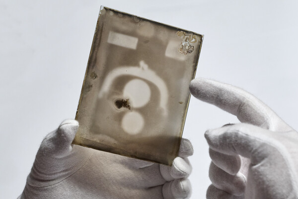
Two X-ray plates from Arthur Goodspeed, believed to have created the world’s first X-ray image, were donated by his family to Penn’s University Archives.