Brain matter altered in U.S. personnel who developed neurological symptoms in Cuba
Brain imaging of 40 U.S. government personnel who experienced a host of neurological symptoms after possible exposure of an unknown source while serving in Cuba revealed significant differences in brain tissue and connectivity when compared to healthy individuals, according to a new report from researchers at the Perelman School of Medicine. The findings are published in JAMA.
The colored regions in these images show differences on the group level—or averages—widespread throughout the brain, and particularly in the cerebellum, which is responsible for balance, coordination, and speech. (Image: JAMA Network)
“The areas implicated in the patients’ brains, namely the cerebellum as well as the visuospatial and auditory networks, align with the neurological symptoms that were observed in the patients,” says lead author Ragini Verma,a professor of radiology and head of the DiCIPHR (Diffusion and Connectomics in Precision Healthcare Research) imaging lab at Penn. “These differences persisted even when people with some history of brain injury were excluded from the analysis.”
In 2016, U.S. government personnel serving in Havana, Cuba, and their family members began to report a variety of neurological symptoms, including difficulty with concentration and memory, dizziness, visual issues, and balance problems. The symptoms were linked to sudden, intensely loud noises heard in their homes and hotel rooms, which State Department officials later referred to as a “sonic attack” or “directional phenomena,” though the specifics of the alleged event remain unsolved. After initial examinations, the patients were sent to Penn’s Center for Brain Injury and Repair for evaluation, treatment, and rehabilitation in the summer of 2017, under the direction of center director and study co-author Douglas H. Smith,the Robert A. Groff Professor of Teaching and Research in Neurosurgery.
The group-based analysis, published in the new 2019 JAMA study, used various computational tools to examine structural, diffusion, and functional MRI images, finding statistically significant differences in brain volume, tissue properties, and connectivity between the patients and healthy control group.
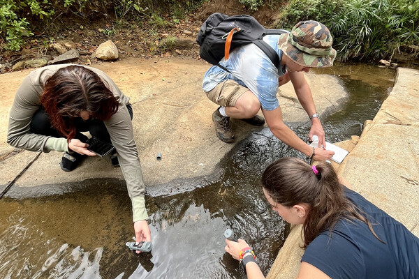
Griffin Pitt, right, works with two other student researchers to test the conductivity, total dissolved solids, salinity, and temperature of water below a sand dam in Kenya.
Griffin Pitt’s upbringing made her passionate about water access and pollution, and Penn has given her the opportunity to explore these issues back home in North Carolina and abroad.
Helping robots work together to explore the Moon and Mars
Penn Engineers, NASA, and five other universities tested robotic systems designed to help unmanned explorers cooperate in the dunes of White Sands, New Mexico, paving the way for Moon and Mars exploration.
From framework to actions: Provost John L. Jackson Jr. talks Penn Forward
In a Q&A, Provost John L. Jackson Jr. explains the relationship between the strategic framework In Principle and Practice and Penn Forward—a new University-wide process and action plan that will advance Penn forward for the next decade and beyond.


