The brain’s amyloid buildup is not a powerful indicator of Alzheimer’s disease
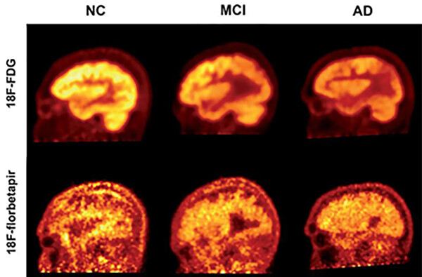
While the presence of beta-amyloid plaques in the brain may be a hallmark of Alzheimer’s disease, giving patients an amyloid PET scan is not an effective method for measuring their cognitive function, according to a new study from researchers in the Perelman School of Medicine and Thomas Jefferson University. The researchers concluded that fluorodeoxyglucose (FDG) PET, which measures the brain’s glucose consumption as a marker of neural activity, is a stronger approach for assessing the progression and severity of Alzheimer’s and mild cognitive impairment (MCI) as compared to florbetapir-PET scans, which reveal amyloid protein deposits in the brain. This suggests that FDG-PET is also a better means for determining the effectiveness of Alzheimer’s therapies, as well as tracking patients’ disease advancement, in both clinical and research settings. Results of this study are detailed in the August issue of the Journal of Alzheimer’s Disease.
PET imaging using the radiotracers FDG and florbetapir to quantify cognitive decline in patients with Alzheimer’s disease (AD), mild cognitive impairment (MCI), and healthy controls. (Image: Penn Medicine News)
“Both florbetapir-PET and FDG-PET are approved diagnostic methods for Alzheimer’s disease, and both appear to be effective in indicating some sort of cognitive impairment. However, we have now shown that FDG-PET is significantly more precise in clinical studies, and it is also available for routine use with modest costs,” says the study’s co-principal investigator Abass Alavi, a professor of radiology at Penn. “Our results support the notion that amyloid imaging does not reflect levels of brain function, and therefore it may be of limited value for assessing patients with cognitive decline.”
Two of the most significant biomarkers found in Alzheimer’s are decreased glucose uptake and the accumulation of amyloid plaques in the brain. PET scans use different radioactive drugs, called radiotracers, to measure these biomarkers within the brain tissue of patients with cognitive impairment. FDG-PET is one of the most commonly used imaging techniques to diagnose Alzheimer’s. However, in recent years, several other radiotracers, such as florbetapir, have been developed to detect the deposition of amyloid plaques.
Recently, the effectiveness of amyloid imaging as a strategy for monitoring dementia symptoms has been called into question.
Griffin Pitt, right, works with two other student researchers to test the conductivity, total dissolved solids, salinity, and temperature of water below a sand dam in Kenya.
Griffin Pitt’s upbringing made her passionate about water access and pollution, and Penn has given her the opportunity to explore these issues back home in North Carolina and abroad.
Helping robots work together to explore the Moon and Mars
Penn Engineers, NASA, and five other universities tested robotic systems designed to help unmanned explorers cooperate in the dunes of White Sands, New Mexico, paving the way for Moon and Mars exploration.
From framework to actions: Provost John L. Jackson Jr. talks Penn Forward
In a Q&A, Provost John L. Jackson Jr. explains the relationship between the strategic framework In Principle and Practice and Penn Forward—a new University-wide process and action plan that will advance Penn forward for the next decade and beyond.


