Colorful research comes to life when everything ‘clicks’ together
A collaborative project connects proteins with fluorescent dyes through azide−alkyne cycloaddition, known as a ‘click’ reaction, that provides researchers with a dynamic glimpse inside living cells.
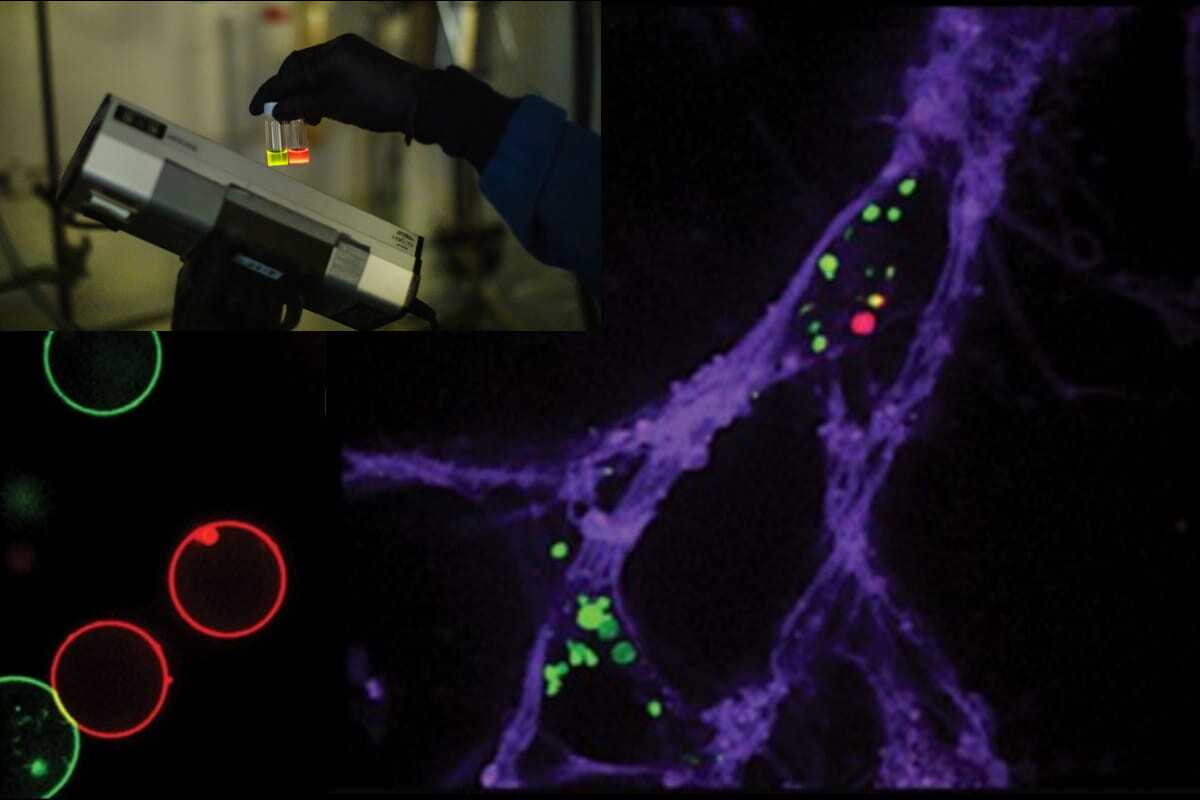
A new fluorescent dye, known as clickable and photoconvertible diazaxanthilidene (CPX for short), changes color when exposed to UV light (left inset) and can be used to track protein aggregates inside neurons (right). (Image: Eric Sucar [left inset], J. Am. Chem. Soc. [right])
After becoming colleagues eight years ago, David Chenoweth and James Petersson quickly found that their overlapping interests made for an ideal collaboration. For both researchers, everything seemed to “click” naturally.
Chenoweth and Petersson are chemical biologists who study molecules that have potential applications in fields like biology and medicine. “But David’s lab is maybe a little bit more capital C and mine is a little more capital B,” explains Petersson. The Chenoweth lab specializes in synthesizing organic molecules while members of the Petersson group are experts in engineering proteins with unique amino acids or fluorescent dyes.
While the groups had worked together previously on several projects, their collaboration officially clicked when they welcomed their first joint graduate student. Joomyung “Vicky” Jun, who earned her undergraduate degree from the University of California, Berkeley, saw the joint position with the Chenoweth and Petersson labs as the perfect place to apply her chemistry knowledge towards making new tools for biologists and medical researchers.
Jun said that working in two labs gave her “a good story” and kept her motivated throughout her Ph.D. making a compound that’s useful for biologists.
Jun worked with a fluorescent dye that can change from green to red when activated with UV light, a molecule that was initially discovered by a graduate student and postdoc in Chenoweth’s lab. Wanting to make good use of this color-changing dye, Jun used techniques developed by the Petersson lab for attaching the dye onto proteins. “We thought it might be easy to just add a linker where we can make this molecule ‘clickable,’ so we can use it for any biomolecule,” explains Jun.
Jun was able to make this dye “clickable” using an azide−alkyne cycloaddition, a chemical reaction that makes it easy to attach the dye molecule onto any protein. The color-changing properties of this new dye, known as clickable and photoconvertible diazaxanthilidene (CPX for short), allows researchers to more easily track how proteins move and change over time while they are inside a cell.
Having colorful, “clickable” chemistry is one thing, but using it on actual living cells is another. To test their new molecule in a biological system, Jun and her mentors turned to the expertise of the group led by Virginia Lee, who has been studying neurodegenerative disease for more than 30 years. Their group’s prior discovery of alpha-synuclein, a protein that forms aggregates in the brain and causes the loss of dopaminergic neurons, led them to finding a way to grow neurons in cell culture so they could more easily study how Parkinson’s disease spreads.
But the methods they could use to study these neuronal cell cultures were limited. “You can fix cells, you can look inside cells and see the pathology, but if you want to observe a live cell you need to develop new ways so you can see what is happening,” says Lee. “The way we decided to do that was by collaborating with James.”
Lee says that Petersson’s creativity and knowledge of chemistry was instrumental in developing this new approach that can now be used to track the movement of alpha-synuclein proteins in real time inside living neuronal cells. “This assay is powerful in that it allows us to look at many different things, and it was made possible through our collaboration,” she says.
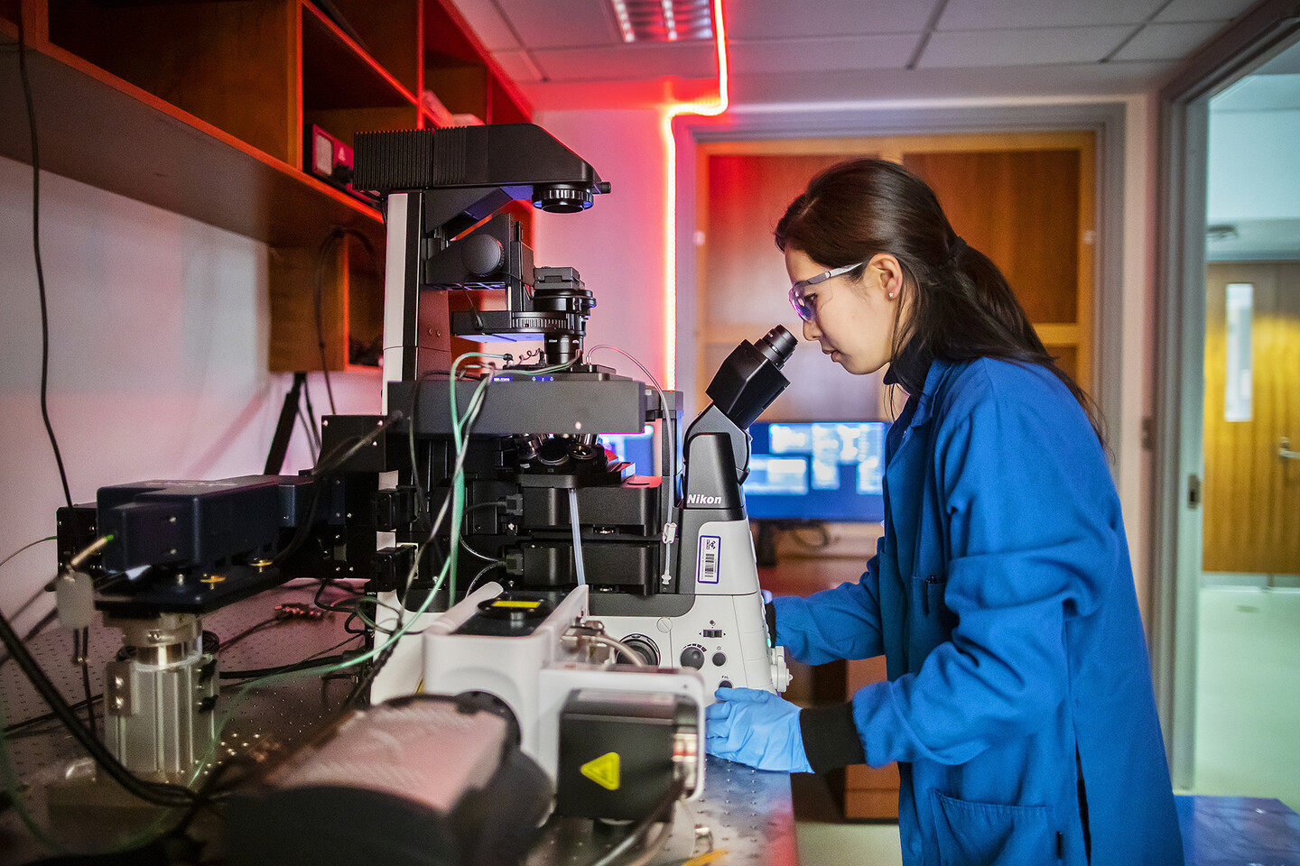
Now, the challenge of collecting images of the CPX-linked alpha-synuclein proteins fell back to the chemists. With the help of engineers at Nikon, Chenoweth lab members designed and built a custom modular microscope for doing photoactivation experiments. From the high-powered lasers that activate CPX’s color-changing abilities to the lenses and mirrors that must be aligned perfectly to capture the images, everything had to come together perfectly. “It was an exciting project for all of us because they had never built this kind of complicated microscope before,” Jun says about the collaboration with the Nikon engineers.
The modular microscope is made of two cameras, two confocal lenses with a high aperture, seven lasers for imaging and three LEDs for stimulating the dye with light, four lasers for photobleaching, and a component that allows the microscope to image the cells using a method known as total internal reflection fluorescence microscopy.
This work was published in the Journal of the American Chemical Society, with collaborative efforts that span chemistry, biology, and medicine being instrumental to its success. “Even if you have a compound, every step needs optimization,” says Jun. “These experiments are not easy, so it has to be highly collaborative. I don’t have enough background for all the biology, but I know what [the biologists] need, and I know what they want to study,” she says.
Chenoweth and Petersson also emphasize the need for these types of cross-collaborative experiments in order to develop the tools and techniques that will take biochemical and medical research to the next level. “It’s something where a lot of your more traditional biochemical experiments are not going to be sufficient,” Petersson says.
“Penn is a really unique place,” says Chenoweth. “We’re lucky in many ways because we have everything here we seem to need between the med school, the dental school, the vet school, and all the engineering departments.”
Lee agrees. “Collaboration benefits everyone,” she says. “If you go to other places, normally they don’t collaborate that much. But here at Penn we have a culture of collaboration, and we facilitate it.”
This research was supported by National Institutes of Health grants R56-NS081033, R01-GM118510, and RR-023444; National Science Foundation Grant MRI 0820996; and funding provided by the School of Arts and Sciences and Dean Steven J. Fluharty.
Nanoparticle blueprints reveal path to smarter medicines
New research involving Penn Engineering shows detailed variation in lipid nanoparticle size, shape, and internal structure, and finds that such factors correlate with how well they deliver therapeutic cargo to a particular destination.
A generous gift from alumni Glenn and Amanda Fuhrman brings the work of internationally acclaimed artist Jaume Plensa to the University of Pennsylvania. The latest addition to the Penn Art Collection expands Philadelphia's public art.
A massive chunk of ice, a new laser, and new information on sea-level rise
For nearly a decade, Leigh Stearns and collaborators aimed a laser scanner system at Greenland’s Helheim Glacier. Their long-running survey reveals that Helheim’s massive calving events don’t behave the way scientists once thought, reframing how ice loss contributes to sea-level rise.


