Imaging just one week after starting treatment can predict melanoma response to immunotherapy
New Penn Medicine research shows that following a single dose of a particular immunotherapy drug, CT scans ‘lit up’ with metabolic changes in tumors that correlated with longer survival.
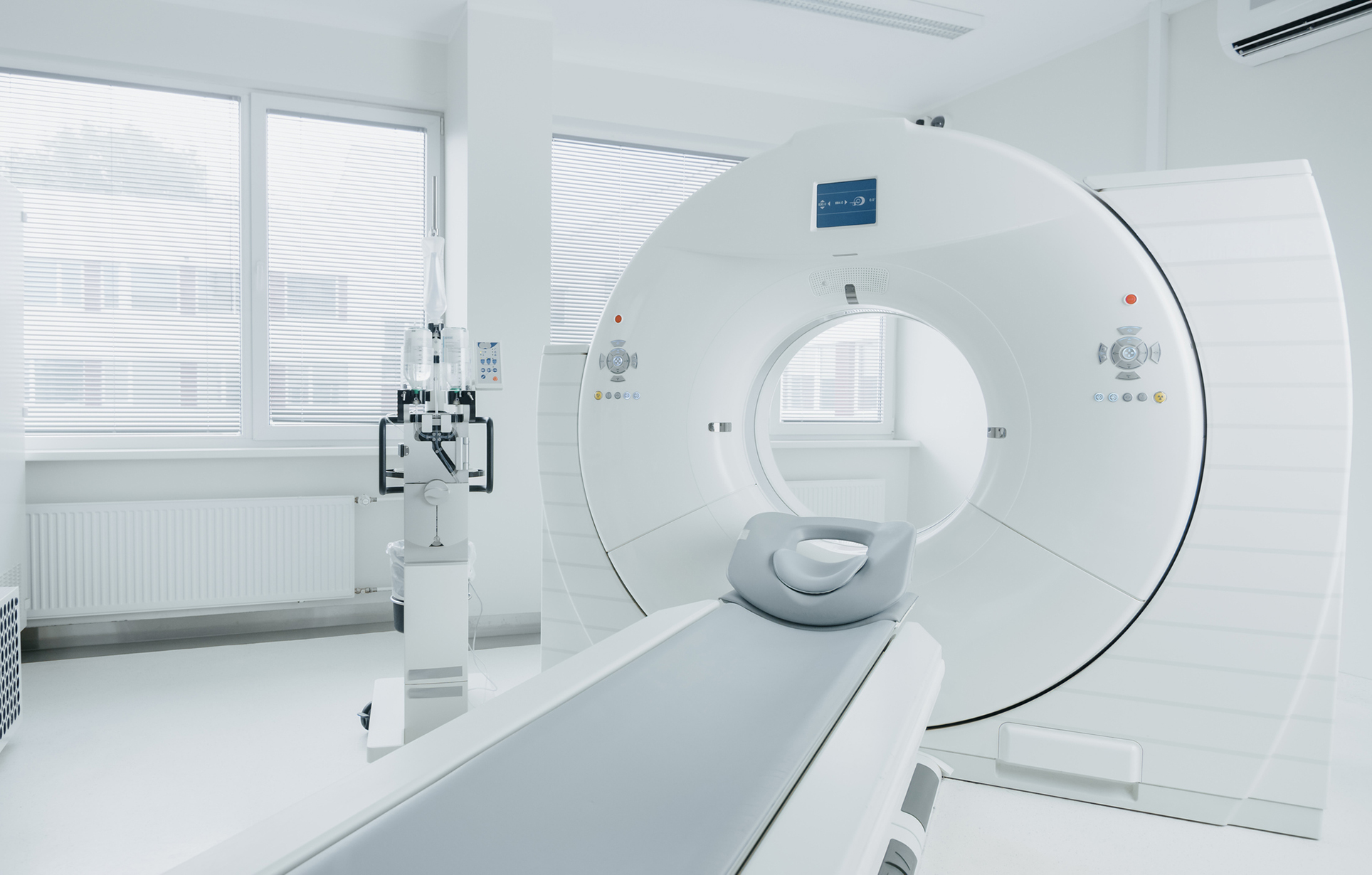
While the standard timing for imaging patients’ tumors after immunotherapy is three months, researchers found that imaging patients with melanoma after just one week of treatment illuminated metabolic changes in their tumors that corresponded with a response to the treatment and longer survival, according to new research from the Perelman School of Medicine.
“Time is always valuable when treating cancer, and imaging tumors early can improve how clinicians develop personalized treatment plans for each patient,” says senior author Michael Farwell, an associate professor of radiology at Penn Medicine. “For example, if a patient’s scan shows a response, they could potentially de-escalate therapy or avoid surgery. On the other hand, if a patient isn’t showing a response, it tells their care team to try other treatment options which could better treat their cancer, and would reduce unnecessary side effects from an ineffective and costly treatment.”
Imaging patients with melanoma after just one week of immunotherapy illuminated metabolic changes in their tumors that corresponded with a response to the treatment and longer survival.
Immunotherapy activates the body’s own immune system to attack tumors. While it can be effective for many patients with cancer, not all patients respond to therapy, and the treatment can cause severe adverse events, like toxicity that affects the stomach and intestine, hormone, or skin.
In the study, published in Clinical Cancer Research, Farwell and his colleagues evaluated whether a 18-F-fluorodeoxyglucose (FDG) positron emission tomography (PET) and computed tomography (CT) scan could detect an increase in metabolic activity in patients shortly after their first dose of immunotherapy. FDG PET and CT is typically used to detect cancer in the body. It is FDG that causes tumors to “light up” in CT and PET scans.
Similarly, when a patient responds to immunotherapy, activated immune cells penetrate the tumor, which shows an increase in FDG activity on scans, which Farwell and his collaborators refer to as a “metabolic flare.” Then, as the tumor responds to therapy, the tumor cells die and pass back through a stable metabolism phase and ultimately show a decrease in FDG activity, which the researchers refer to as a state of “metabolic response.” In contrast, the tumors of nonresponding patients maintain stable metabolism.
In Senegal, the ambitious Dakar Greenbelt project seeks to create an extensive network of ecological infrastructure in and around the city to sustainably address environmental concerns and enhance urban life. With support from David Gouverneur and Ellen Neises, Ph.D. candidate Rob Levinthal in the Weitzman School of Design led two courses that included a field trip to Dakar, that culminated in students presenting their visions for parts of the Greenbelt.
From a desert to an oasis: Penn engages in ambitious greening effort in the Sahel
Students from the Weitzman School of Design journeyed to Senegal to help with a massive ecological and infrastructural greening effort as part of their coursework. The Dakar Greenbelt aims to combat desertification and promote sustainable urban growth.
As part of an undergraduate course, Penn faculty and students curated an Arthur Ross Gallery exhibition of works from the Neumann family’s extensive collection of modern and contemporary art.
The University’s nexus for technology transfer supports researchers in their innovative efforts, from CAR T to mRNA advancements that have dramatically reshaped the world.