(From left) Doctoral student Hannah Yamagata, research assistant professor Kushol Gupta, and postdoctoral fellow Marshall Padilla holding 3D-printed models of nanoparticles.
(Image: Bella Ciervo)
2 min. read
Cell replacement therapy offers new hope for millions of people affected by retinal degenerations (RDs)–a group of blinding conditions caused by the loss of light-sensing photoreceptor cells in the retina. Among the most promising approaches is the transplantation of stem cell-derived partially differentiated photoreceptor cells, known as precursor cells, to replace those lost to disease. But a persistent hurdle remains: many of the transplanted cells do not survive long enough to integrate or restore vision.
Now, researchers from the Division of Experimental Retinal Therapies (ExpeRTs) at Penn’s School of Veterinary Medicine, led by Raghavi Sudharsan, and William A. Beltran, in collaboration with researchers at the University of Wisconsin-Madison and Harvard Medical School, have uncovered a key reason for this early transplant failure. In a study published in Stem Cell Research & Therapy, the team reports that photoreceptor precursor cells experience widespread death within the first few days after being injected into the subretinal space, even under conditions of effective immune suppression. The culprit, they find, is acute metabolic stress triggered by a sudden shift from a nutrient-rich culture environment into the relatively nutrient-deprived conditions of the subretinal space—a transition that triggers rapid cell loss during the first few days after transplantation. The study points to a critical need for strategies that help donor cells survive this abrupt metabolic transition and better adapt to the challenging conditions of the host retina.
Using noninvasive imaging, the researchers find that early donor cell death is consistently observed, regardless of whether the host retina is healthy or already undergoing degeneration. To uncover the underlying cause, they performed single-cell RNA sequencing on transplanted cells in healthy retinas, which reveals signatures of acute metabolic stress.
The study reveals a positive aspect: A subset of donor cells did survive and continued to mature after transplantation. In retinas that retained some of the native photoreceptors, these surviving cells were even able to begin forming structures that resembled synaptic connections. In models where portions of the retina’s photoreceptor layer were still intact, donor cells could integrate into the host tissue. But in cases of end-stage degeneration, where the retinal architecture was too far gone, transplanted cells failed to survive.
“As the field moves closer to clinical translation,” says Sudharsan, “understanding how both the transplanted cells and the host retina respond will be essential to designing therapies that actually succeed in patients.”
This story is by Raghavi Sudharsan and William A. Beltran. Read more at Penn Vet News.
From Penn Vet

(From left) Doctoral student Hannah Yamagata, research assistant professor Kushol Gupta, and postdoctoral fellow Marshall Padilla holding 3D-printed models of nanoparticles.
(Image: Bella Ciervo)

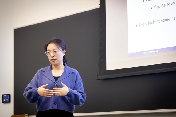
Jin Liu, Penn’s newest economics faculty member, specializes in international trade.
nocred

nocred

nocred