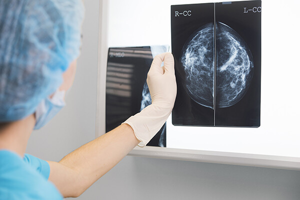
Penn researchers can predict 10-year breast cancer recurrence with MRI scans that characterize the genetic makeup of tumors.
Diverse diseases like breast cancer can present challenges for clinicians, specifically on a cellular level. While one patient’s tumor may differ from another’s, the cells withinthetumor of a single patientcan also vary greatly. This can be problematic, considering that an examination of a tumor usually relies on a biopsy, which only captures a small sample of the cells.
According to a new study from researchers at Penn Medicine, magnetic resonance imaging (MRI) and an emerging field of medicine called radiomics—which uses algorithms to extract a large amount of features from medical images—could help to characterize the heterogeneity of cancer cells within a tumor and allow for a better understanding of the causes and progression of a person’s individual disease. The findings were published in Clinical Cancer Research.
“If we’re only taking out a little piece of a tissue from one part of a tumor, that does not give the full picture of a person’s disease and of his or her response to specific therapies,” says principal investigator Despina Kontos,an associate professor of radiology in the Perelman School of Medicine. “We know that in a lot of instances, patients are over-treated, getting therapy that may not be beneficial. Or, conversely, patients who need more aggressive therapy may not end up receiving it. The method we currently have for choosing the appropriate treatment for patients with breast cancer is not perfect, so the more steps we can take toward more personalized treatment approaches, the better.”
Nanoparticle blueprints reveal path to smarter medicines
New research involving Penn Engineering shows detailed variation in lipid nanoparticle size, shape, and internal structure, and finds that such factors correlate with how well they deliver therapeutic cargo to a particular destination.
A generous gift from alumni Glenn and Amanda Fuhrman brings the work of internationally acclaimed artist Jaume Plensa to the University of Pennsylvania. The latest addition to the Penn Art Collection expands Philadelphia's public art.
A massive chunk of ice, a new laser, and new information on sea-level rise
For nearly a decade, Leigh Stearns and collaborators aimed a laser scanner system at Greenland’s Helheim Glacier. Their long-running survey reveals that Helheim’s massive calving events don’t behave the way scientists once thought, reframing how ice loss contributes to sea-level rise.


