Sheeba never strays far from her owner John Vagner’s side. She’s his best bud and, as a diabetic alert dog trained by nonprofit Canine Partners for Life, his most astute signaler of a blood sugar drop or spike. The Labrador’s keen senses, particularly sight and smell, help her be the hard-working, loyal partner she is.
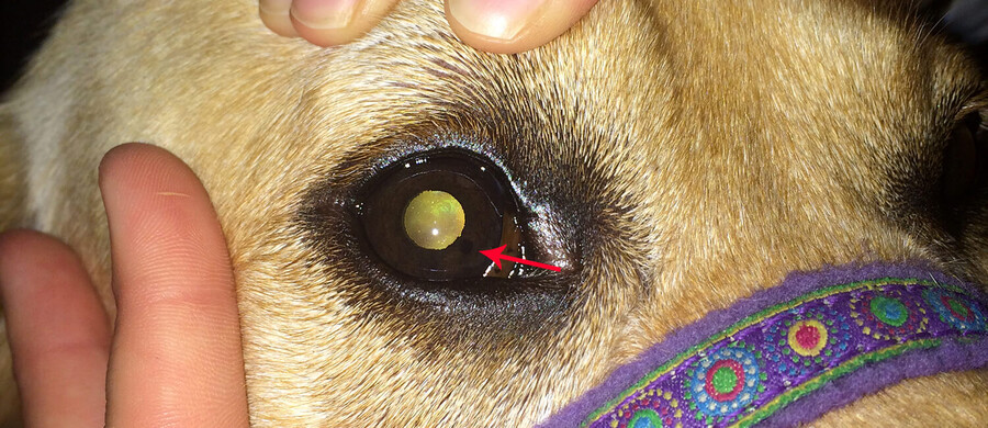
Sheeba’s lesion, the black dot at the red arrow, can be seen by the naked eye. (Photo: Penn Vet News)
Every May, Vagner takes Sheeba for an eye check-up during the National Service Dog Eye Exam, an annual event sponsored by the American College of Veterinary Ophthalmologists (ACVO). The Ryan Veterinary Hospital is a regular participant in the ACVO program, offering eye exams to screen for conditions that could jeopardize a working dog’s vision.
“We have a technique called ultrasound biomicroscopy—or UBM,” says Keiko Miyadera, assistant professor of ophthalmology. “It’s a noninvasive high-resolution imaging of the eye that enables us to pick up details we wouldn’t see in regular screening exams.” Using the technology, Miyadera measured a 1.7 millimeter lesion on the iris in Sheeba’s right eye.
Since 2015, Miyadera has examined Sheeba’s eye once a year. And every time the lesion’s margins have grown insignificantly. Until 2018.
“I measured a bit more growth during last year’s exam,” Miyadera explained. “So, we went ahead with laser therapy to reduce the size of the mass and decrease the change of further growth.”
Materials in the Annenberg School for Communication Library Archives include thousands of TV scripts, the first issue of TV Guide, and interviews about the early days of HBO—which help to chronicle TV’s 100-year story.
Centering joy in AI development and implementation
PIK Professor Desmond Upton Patton—of Annenberg and SP2—and collaborators introduce a joy-informed framework designed to initiate conversations among engineers, designers, and researchers.
Winter Storm Fern brought icy and snowy conditions to the Northeast and other parts of the country over the weekend. Penn Today asks physicist Robert Carpick about the unique properties of ice, the science of curling, and how close we are to ‘nonslip’ ice.
Organizations like Penn’s Netter Center for Community Partnerships foster collaborations between Penn and public schools in the West Philadelphia community.
Penn receives national distinction for community engagement
The recognition by the American Council on Education and Carnegie Foundation for the Advancement of Teaching acknowledges Penn’s long-standing commitment to community-engaged scholarship and partnerships in West Philadelphia and beyond.


