Latest ‘organ-on-a-chip’ is a new way to study cancer-related muscle wasting
Studying drug effects on human muscles just got easier thanks to a new “muscle-on-a-chip,” developed by a team of researchers from Penn’s School of Engineering and Applied Science and Inha University in Incheon, Korea.
Bioengineering’s Dan Huh and colleagues have developed a number of organ-on-a-chip devices to simulate how human cells grow and perform in their natural environments. Their latest is a muscle-on-a-chip, which carefully captures the directionality of muscle cells as they anchor themselves within the body. (Image: Melissa Pappas).
Muscle tissue is essential to almost all of the body’s organs, however, diseases such as cancer and diabetes can cause muscle tissue degradation or “wasting,” severely decreasing organ function and quality of life. Traditional drug testing for treatment and prevention of muscle wasting is limited through animal studies, which do not capture the complexity of the human physiology, and human clinical trials, which are too time consuming to help current patients.
An “organ-on-a-chip” approach can solve these problems. By growing real human cells within microfabricated devices, an organ-on-a-chip provides a way for scientists to study replicas of human organs outside of the body.
Using their new muscle-on-a-chip, the researchers can safely run muscle injury experiments on human tissue, test targeted cancer drugs and supplements, and determine the best preventative treatment for muscle wasting.
This research was published in Science Advances and was led by Dan Huh, associate professor in the Department of Bioengineering, and Mark Mondrinos, then a postdoctoral researcher in Huh’s lab and currently an assistant professor of biomedical engineering at Tulane University.
“Using the chip in conjunction with a computational model, we found that when mechanical stress was generated in opposing directions during the sculpting of muscle tissue, the contractility, or the ability of a cell to contract, increased in the direction it was being pulled. This caused the alignment and stiffening of the extracellular matrix in that direction, which then signaled the cells to further increase their contractility,” says Huh. “Creating the 3D muscle tissue on the chip was key in discovering this new feedback mechanism.”
The muscle chip was used for studying more than just the size, shape and strength of the cultured muscle cells.
Deepfakes, digital doubles, and the law: Jennifer Rothman on protecting identity in the AI era
The evolution of AI technology may require more robust copyright laws and guardrails that protect people’s control of their own identities, says the Penn Carey Law professor.
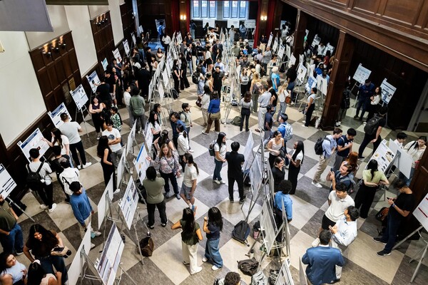
Bold ideas and innovation on display at the Fall Research Expo
On Sept. 15, hundreds of posters were presented throughout Houston Hall at the annual Fall Research Expo, representing the research projects of 410 undergraduate students conducted through the Center for Undergraduate Research.
Exposure to air pollution worsens Alzheimer’s disease
New research from Penn Medicine finds living in areas with high concentration of air pollution is associated with increased buildup of amyloid and tau proteins in the brains of Alzheimer’s patients, accelerating cognitive decline.


