Researchers from the School of Arts & Sciences have shown that bacteria from extreme marine environments can help reduce asbestos’ toxic properties.
Asbestos materials, a group of naturally occurring minerals once widely used in a range of industries for their strength and heat resistance, are notorious for being a health hazard. While their use has declined substantially, the minerals are not banned in the United States, and people can still be exposed when asbestos-containing buildings are disturbed during renovations or demolition.
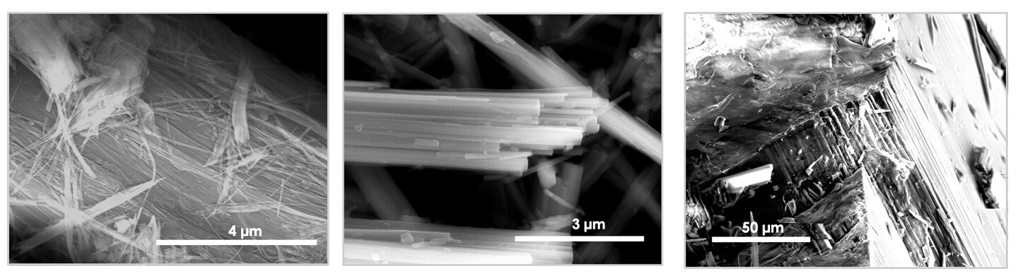
Minerals used in the study, from left to right: chrysotile, crocidolite, and tremolitic actinolite.
(Images: Ruggero Vigliaturo)
The hazard hit close to home when several schools in the Philadelphia school district were closed after failing asbestos inspections. Penn has pledged a $100 million contribution to the School District—$10 million annually for 10 years—to be used to remediate environmental hazards, including asbestos and lead, in public school buildings. Better remediation options, however, are needed for dealing with asbestos.
Now, researchers from Penn’s Department of Earth and Environmental Science show that bacteria from extreme marine environments have the potential to detoxify asbestos. Their study, published in the journal Applied and Environmental Microbiology, suggests that the marine microbes may be better candidates for asbestos bioremediation than previously tested fungi and soil bacteria.
“We wanted to expand asbestos bioremediation research by exploring ways to lower the toxicity of these minerals for safer disposal or reuse as secondary raw materials,” says senior author Ileana Pérez-Rodríguez, an assistant professor of earth and environmental science who specializes in studying extremophilic deep-sea microbes.
To do this, Pérez-Rodríguez teamed with Reto Gieré, who has a long history characterizing asbestos minerals. They thought that these extremophilic microbes might be good candidates for asbestos bioremediation because they use inorganic compounds and interact with a variety of minerals in their natural environments.
Senior author Ileana Pérez-Rodríguez (right) teamed with Reto Gieré to study the potential for two bacterial species, Deferrisoma palaeochoriense and Thermovibrio ammonificans, to detoxify asbestos.
(Images: Courtesy of Reto Gieré and Brooke Sietinsons)
The team focused on two bacterial species, Deferrisoma palaeochoriense and Thermovibrio ammonificans, to target two aspects of asbestos minerals that make them dangerous when inhaled: their iron content, which is largely responsible for the material’s carcinogenic effects, and their fibrous structure, which causes inflammation.
To test the microbes’ ability to detoxify asbestos, the researchers incubated them for seven days at 60 °C or 75 °C—the microbes’ preferred temperatures—in small liquid-filled bottles that also contained asbestos minerals. Across this period, the researchers took samples of the liquid media to track cell growth and changes in chemical composition, and they used electron microscopy to look for changes in mineral structure.
They found that D. palaeochoriense, which uses iron as part of its metabolism, could effectively remove some iron from asbestos while using it to grow. However, this removal of iron did not change the mineral’s overall fibrous structure, which is partially responsible for its toxicity.
“This is a gradual process of taking a highly hazardous mineral and making it less hazardous,” Pérez-Rodríguez says. “You can make the mineral less toxic by eliminating the chemical reactivity that comes with the iron, but you still have that fibrous structure, so the next question is, ‘How do we break down the shape?’”
Asbestos minerals are composed of a silicate backbone, and previous studies have shown that removing silicon and magnesium ions from this backbone can disrupt its fibrous structure. This is where the second bacteria, T. ammonificans, came in.
“We can see through microscopy that these microbes incorporate silicon into their biofilms” Pérez-Rodríguez says. “Usually when we think about biofilms, we think about a sort of slimy goo, but in this case the biofilms are actually quite rigid; they’re basically creating little houses made out of rocks.”
Tubular structures resembling “small vent chimneys” were observed in experimental titanium foil surfaces provided to deep-sea vent bacterium T. ammonificans while growing in the presence of chrysotile asbestos for seven days.
(Images: Jessica Choi)
The researchers found that T. ammonificans could accumulate silicon from “serpentine” asbestos, which has curly fibers, but not from “amphibole” asbestos, which has straight, needle-shaped fibers. “This really highlights the difficulty of approaching asbestos treatments as a one-size-fits-all solution, given the unique chemical compositions and crystal structures associated with each asbestos mineral,” Pérez-Rodríguez says.
Microbial-based asbestos treatments are a desirable alternative to current asbestos treatment methods which involve either heating it to very high temperatures and pressures or by treating it with strong acids or bases. However, more research is needed to test how these methodologies could be used to remediate asbestos on a large scale.
“This was just a first lab test, and of course there are still questions, and we would have to do much more research, but hopefully we can take it to the next level,” Gieré says.
The study was co-authored by former postdoctoral researchers Jessica Choi, now a postdoctoral researcher at the University of Michigan, and Ruggero Vigliaturo, now an assistant professor at the University of Turin.
This research was supported by Penn Start-up Funds, a Penn Elliman Faculty Fellowship and the Penn Center of Excellence in Environmental Toxicology (Grant P30-ES013508), as well as by the National Institute of Environmental Health Sciences of the National Institutes of Health (P42-ES023720). Part of this work was carried out at the Penn’s Singh Center for Nanotechnology, part of the National Nanotechnology Coordinated Infrastructure Program, which is supported by National Science Foundation (Grant NNCI-1542153).
Materials in the Annenberg School for Communication Library Archives include thousands of TV scripts, the first issue of TV Guide, and interviews about the early days of HBO—which help to chronicle TV’s 100-year story.
Centering joy in AI development and implementation
PIK Professor Desmond Upton Patton—of Annenberg and SP2—and collaborators introduce a joy-informed framework designed to initiate conversations among engineers, designers, and researchers.
Winter Storm Fern brought icy and snowy conditions to the Northeast and other parts of the country over the weekend. Penn Today asks physicist Robert Carpick about the unique properties of ice, the science of curling, and how close we are to ‘nonslip’ ice.
Organizations like Penn’s Netter Center for Community Partnerships foster collaborations between Penn and public schools in the West Philadelphia community.
Penn receives national distinction for community engagement
The recognition by the American Council on Education and Carnegie Foundation for the Advancement of Teaching acknowledges Penn’s long-standing commitment to community-engaged scholarship and partnerships in West Philadelphia and beyond.


