Uncovering the role of skin microbiome and immune response in cutaneous leishmaniasis
Two new studies led by Phillip Scott of the School of Veterinary Medicine and Elizabeth Grice of the Perelman School of Medicine demonstrate how bacteria found in leishmaniasis skin lesions and an associated immune response drive disease burden and treatment failure—and suggest new possibilities for treatment of the parasitic disease.
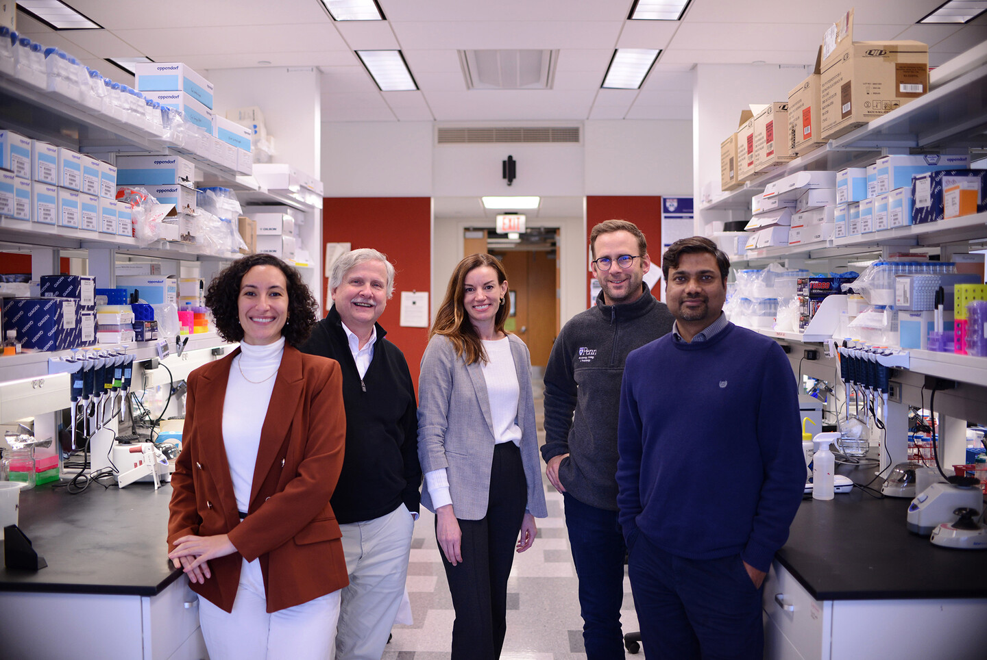
Researchers at Penn’s School of Veterinary Medicine and the Perelman School of Medicine studying leishmaniasis are leading the way to potential new therapies. From left, Camila Amorim, Phillip Scott, Elizabeth A. Grice, Daniel P. Beiting, and Tej Singh.
Image: John Donges for Penn’s School of Veterinary Medicine.
The parasitic disease leishmaniasis is found primarily in Central and South America, the Mediterranean region, the Middle East, and Central Asia and is transmitted to humans by a sand fly. An estimated 1.5 million new cases of cutaneous leishmaniasis—the most common form of the disease—occur each year. There is no vaccine, and treatment with anti-parasitic drugs is often unsuccessful.
People with cutaneous leishmaniasis can develop painful and disfiguring skin ulcers that, if left untreated, may metastasize to the nasopharyngeal region and turn into mucosal leishmaniasis, a more debilitating version of the disease that is untreatable.
Now, two new studies led by researchers at the University of Pennsylvania’s School of Veterinary Medicine and the Perelman School of Medicine have shed light on the complex pathology of the parasitic disease leishmaniasis, pointing the way to potential new therapies.
“It’s a horrific disease,” says Phillip Scott, vice dean of Penn Vet and a longtime leishmaniasis researcher. Some years ago, Scott started collaborating with Dan Beiting, associate professor in Penn Vet's Department of Pathobiology to look at the transcriptional profile of the human disease. This work was expanded to investigate the role of bacteria in the skin, and Beiting established a collaboration with Elizabeth A. Grice, an associate professor in Perelman’s department of dermatology and an expert on the skin microbiome—the microorganisms on the skin. “We started collaborating because we realized that the skin microbiome might be influencing what's happening with the disease,” Scott says.
It turns out they were right.
With the support of researchers in Brazil, they uncovered the pivotal role of the host’s immune response to leishmaniasis, a pathologic response influenced by the presence of bacteria in skin lesions.
The researchers found that it’s often the host’s immune response to parasitic infection, rather than the parasite burden itself, that promotes the most severe disease.
“You need a drug that not only controls the parasite,” Scott says, “but, also one that limits a pathologic host inflammatory response. And so, we're very interested in trying to downregulate that inflammatory response to lessen disease.”
Scott and Grice also show that the skin microbiome plays a significant role in promoting those immunopathologic responses. Using a combination of transcriptional analysis and 16S sequencing in lesions from patients, they found that high levels of bacteria in those lesions—specifically, high levels of Staphylococcus aureus—are associated with treatment failure. These results suggest that therapies that reduce the S. aureus burden in leishmaniasis lesions might promote more rapid healing.
“People have been studying leishmaniasis for many years, and they've thought of it as being caused by one pathogen. It's not,” says Scott “It is a disease caused both by the parasite and the increased bacterial burden in lesions.”
Grice says, “The important takeaway here is that the microbiome can modify the severity of the infections and influence the disease outcomes. Our approach of studying human leishmania infections to identify markers of disease severity, and then taking those findings into model systems to study mechanisms is very powerful.”
To understand the mechanisms involved in the increased pathology associated with bacteria, the researchers studied S. aureus infections in mice with leishmaniasis. They found that colonization of the skin with S. aureus led not only to more severe disease, but also to an increase in regulatory T cells, or Tregs. These cells are known to suppress immune responses and play a critical role in preventing autoimmunity.
To determine the role of Tregs in leishmaniasis, the researchers partially deleted them from the mouse models and found that those mice exhibited more severe disease compared to the mice with normal Treg numbers. Importantly, this increased disease was not associated with more parasites, but was associated with a large increase in the S. aureus burden.
To find out if the results in mice held true for human patients, they returned to their transcriptional analysis of human leishmaniasis lesions and found variability in Tregs within those lesions, as assessed by expression of the Treg transcription factor FOXP3. Significantly, they found that patients with a low expression of FOXP3 in lesions, suggesting fewer Tregs, exhibited delayed healing compared with patients with a high expression of FOXP3.
Unexpectedly, the researchers also found that IFN-gamma, a cytokine normally associated with protection in leishmaniasis, was elevated in patients who exhibited delayed healing. These results show that IFN-gamma can play a pathologic role in leishmaniasis when lesions are co-infected with S. aureus, and provide the foundation for new studies to uncover how IFN-gamma promotes more severe disease.
Scott says the investigators’ findings carry a message. “When studying infectious diseases, we should study them understanding that there are often multiple pathogens involved,” he says. “This is well understood in the gut but is also occurring in the skin.”
The research has important implications for leishmaniasis treatment.
Scott notes that these results indicate that curing leishmaniasis will not only involve killing the parasite with drugs but also controlling a S. aureus infection. We never realized this before. But it’s important, since S. aureus can spread and cause life-threatening infections.
To Grice, the research on leishmaniasis is important—and urgent. “I think about it in terms of helping a population that's burdened with this disease,” she says. But she anticipates that population will grow: With the reality of climate change, the sand fly is expected to expand its geographical habitat. “This is going to affect a larger part of the global population,” says Grice.
Add in the fact that many bacteria, including S. aureus, are becoming multi-drug resistant, she adds, and the need to identify alternative therapies becomes more pressing.
The researchers’ collaboration is typical for Penn, Scott says: “It's just another example of how faculty from different schools at Penn collaborate, it's one strong biomedical community. It is the strong collaborations between faculty that makes Penn such a great place to be.”
Phillip Scott is vice dean for research and academic resources and a professor of microbiology and immunology in the Department of Pathobiology in the University of Pennsylvania School of Veterinary Medicine. He is corresponding author on the Journal of Experimental Medicine paper and co-corresponding author on the Science Translational Medicine paper.
Elizabeth A. Grice is the Sandra J. Lazarus Associate Professor in Dermatology at the University of Pennsylvania’s Perelman School of Medicine’s Penn Institute for Immunology. She is corresponding author on the Journal of Experimental Medicine paper and co-corresponding author on the Science Translational Medicine paper.
Scott and Grice coauthored the studies with researchers from Penn Vet and the Perelman School of Medicine, including lead authors Camila Amorim and Tej Pratap Singh, and co-authors Victoria Lovins, Charles Bradley, Jordan Harris, Fernanda Novais, and Dan Beiting. Critical for this work were collaborators from the Universidade Federal da Bahia in Brazil, including Alexsandro Lago, Lucas Carvalho and Edgar Carvalho.
This work was funded by the National Institutes of Health (R01AI143790, R01NR015639, R01AI162711, P50AI030639, and RO1AI149456), the Penn Skin Biology and Disease Resource–based Center (Penn SBDRC supported by NIH/NIAMS P30AR069589), the National Institute of Arthritis and Musculoskeletal and Skin Diseases (NIAMS F31AR079901 and F31AR079845 and the Penn Dermatology Research T32 Training Grant NIH/NIAMS (T32AR007465).
Nanoparticle blueprints reveal path to smarter medicines
New research involving Penn Engineering shows detailed variation in lipid nanoparticle size, shape, and internal structure, and finds that such factors correlate with how well they deliver therapeutic cargo to a particular destination.
A generous gift from alumni Glenn and Amanda Fuhrman brings the work of internationally acclaimed artist Jaume Plensa to the University of Pennsylvania. The latest addition to the Penn Art Collection expands Philadelphia's public art.
A massive chunk of ice, a new laser, and new information on sea-level rise
For nearly a decade, Leigh Stearns and collaborators aimed a laser scanner system at Greenland’s Helheim Glacier. Their long-running survey reveals that Helheim’s massive calving events don’t behave the way scientists once thought, reframing how ice loss contributes to sea-level rise.


