Leveraging the body’s postal system to understand and treat disease
An interdisciplinary team of researchers in the School of Engineering and Applied Science, Perelman School of Medicine, and School of Arts & Sciences has developed a technique that allows for characterization of both individual carrier and cargo for clinically important molecules.
A research team led by Jina Ko of the School of Engineering and Applied Science and Perelman School of Medicine has developed a new way to characterize the contents of a cargo-carrying particle excreted by many cells, extracellular vesicles.
(image: iStock / Love Employee)
Akin to the packages sent from one person to another via an elaborate postal system, cells send tiny parcels that bear contents and packaging material that serve key purposes: To protect the contents from the outside world and to make sure it gets to the right place via a label with an address.
These packages are known as extracellular vesicles (EVs)—lipid-bound molecules that serve a variety of regulatory and maintenance functions throughout the body. They assist in the removal of unwanted materials within the cell, and they transport proteins, aid in DNA and RNA transfer, and promote tumorigeneses in cancerous cells.
Given their myriad roles, EVs have taken center stage for many researchers in the biomedical space as they have the potential to improve current methods of disease detection and treatment. The main challenge, however, is accurately identifying the molecular contents of EVs while also characterizing the EVs, which, unlike other cellular components that are more homogenous, have more heterogeneity.
Now, a team of researchers at the University of Pennsylvania has developed a novel platform, droplet-free double digital assay, for not only profiling individual EVs but also accurately discerning their molecular contents. The researchers took the digital assay, which quantifies the contents of a molecule via binary metric—a 1 corresponds to the presence of a molecule and a zero to the lack thereof—and applies it to the EV. The work is published in Advanced Science.
The team was led by Jina Ko, an assistant professor with appointments in the School of Engineering and Applied Science and Perelman School of Medicine. “Our method allows for highly accurate quantification of the individual molecules inside an EV,” Ko says . “This opens up many doors in the realm of early disease detection and treatment.”
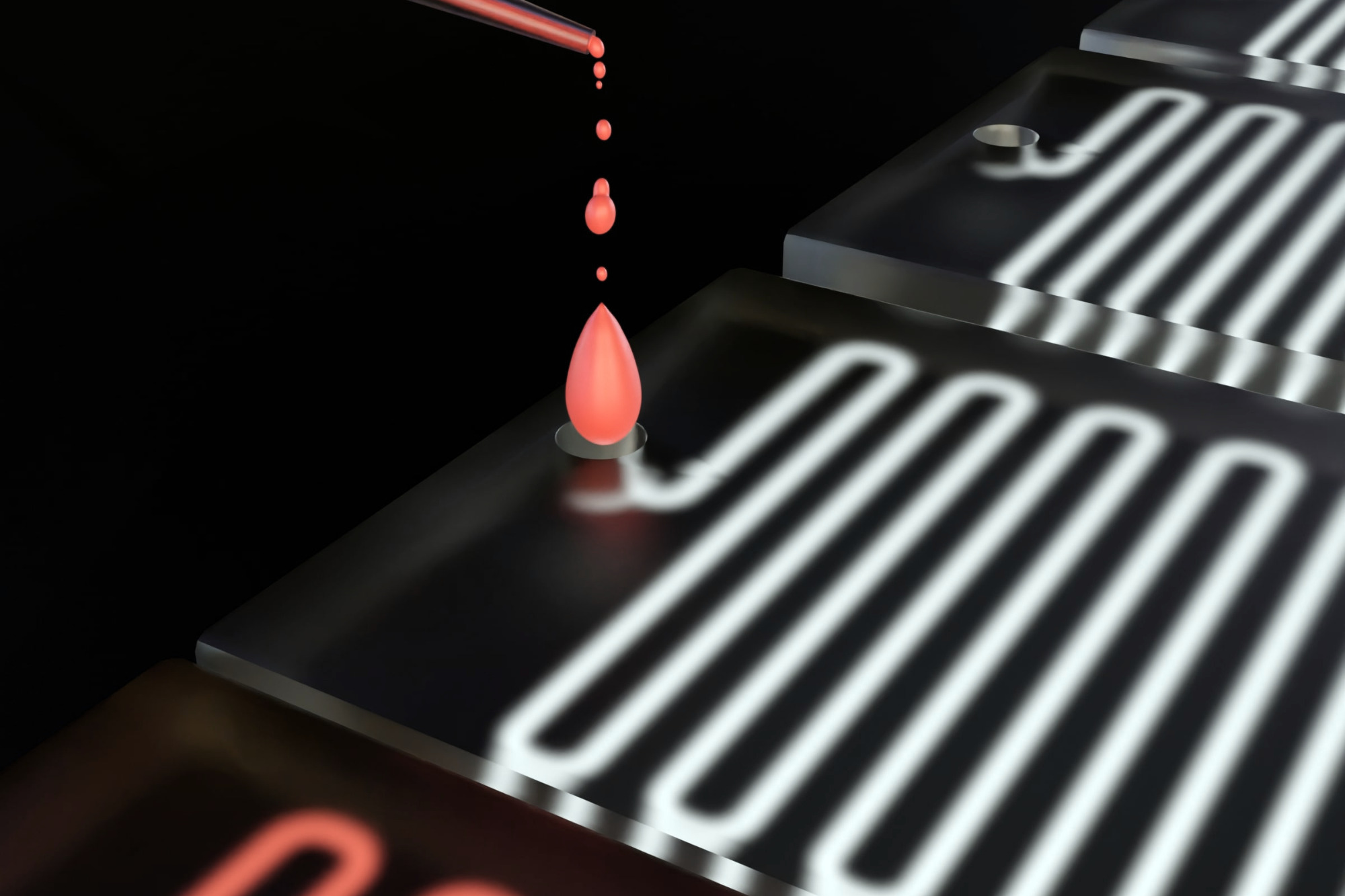
The researchers first compartmentalized individual EVs utilizing a microwell approach to isolate the EVs. Next, they captured individual molecules within the EVs and amplified the signal for clarity. The team then was able to determine the expression levels of pivotal EV biomarkers with remarkable precision via fluorescence.
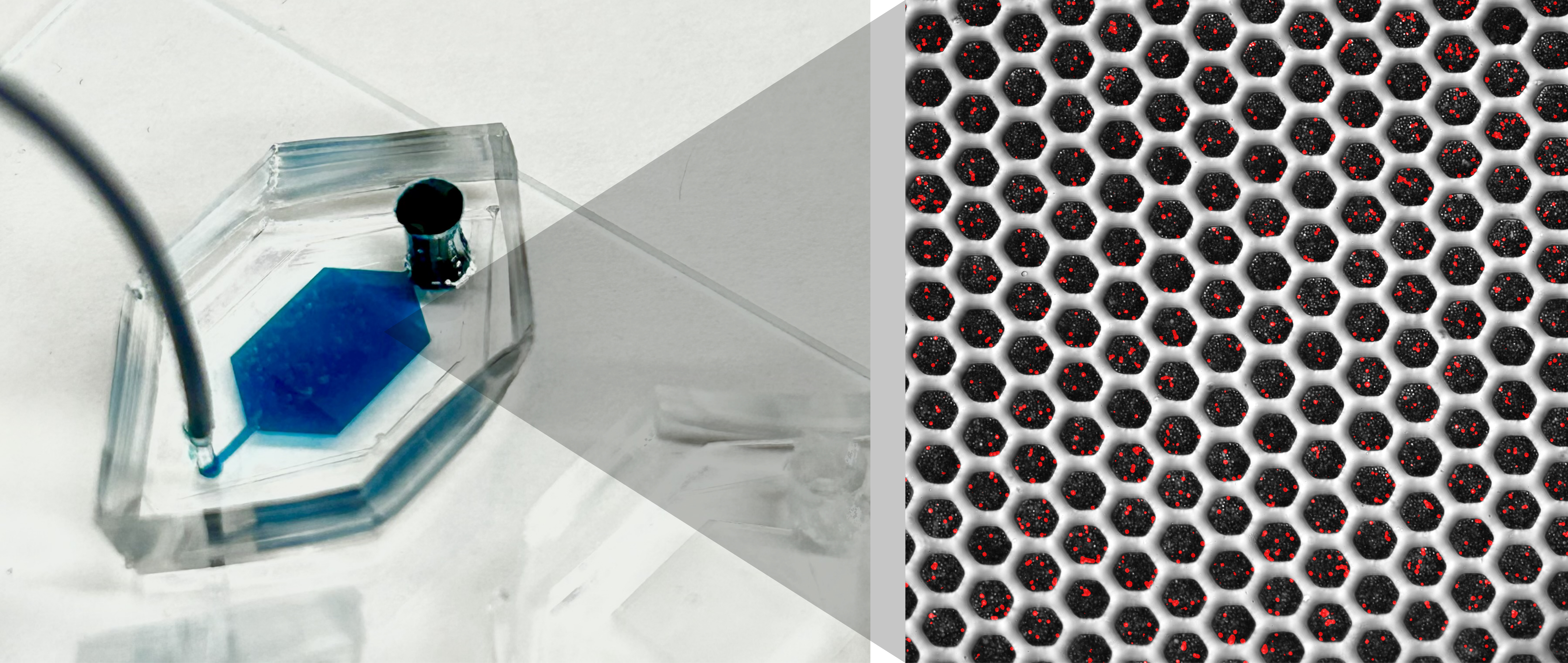
Microwell device with a solution in the reservoir (left) and a visualization of beads capturing extracellular vesicle contents (right).
(Image: Courtesy of David E. Reynolds)
The technique was first tested by partitioning EVs from cancerous tissue samples and characterizing the expression of epidermal growth factor receptor, a protein that plays a key role in cell growth and differentiation, by using signal amplification. Following this, the method was used to determine the abundance of the programmed death-ligand 1 (PD-L1) protein in melanoma cell lines in collaboration with Wei Guo in the Department of Biology at Penn. The results showcased the system's accuracy and sensitivity in detecting EV cargo.
Bioengineering Ph.D. candidate David Reynolds, the paper’s first author says, “PD-L1 molecular detection is particularly useful in cancer research, so it’s exciting to see that the technology we’ve developed could be used to answer some fundamental biology questions and also be translated to an active area of clinical research.”
The researchers note that PD-L1 is a protein that plays a crucial role in regulating the immune system. Specifically, it can suppress the immune response, which under normal conditions helps the body prevent autoimmune reactions but in many cancer cells can be upregulated as a mechanism to “hide” from the immune system.
“Successfully testing our technology with PD-L1 detection hints at the promise of better biomarkers for monitoring tumor behavior, growth, and therapy response,” Ko says. “Looking ahead, we’re excited to explore analytical tools that will provide means to improve our understanding of other cellular components and processes that can tell us more about what our bodies are up to.”
Ko says, "The development of ultra-sensitive tools that can measure clinically important biomarkers with high accuracy will open doors to a new era in biomedical research and its practical applications."
Jina Ko is an assistant professor in the Department of Pathology and Laboratory Medicine in the Perelman School of Medicine and an assistant professor in the Department of Bioengineering in the School of Engineering and Applied Science at the University of Pennsylvania.
David Reynolds is a Ph.D. candidate in the Department of Bioengineering in Penn Engineering.
Other authors include, Menghan Pan, George Galanis, Yoon Ho Roh, Renee-Tyler T. Morales, Shailesh Senthil Kumar, and Su-Jin Heo of the Department of Bioengineering at Penn Engineering; Jingbo Yang and Xiaowei Xu of the Department of Pathology and Laboratory Medicine at Penn Medicine; and Wei Guo of the Department of Biology in the School of Arts & Sciences at Penn.
The research was supported by the National Institutes of Health: grants R00CA256353, R35 GM141832, and CA174523 (SPORE).
In Senegal, the ambitious Dakar Greenbelt project seeks to create an extensive network of ecological infrastructure in and around the city to sustainably address environmental concerns and enhance urban life. With support from David Gouverneur and Ellen Neises, Ph.D. candidate Rob Levinthal in the Weitzman School of Design led two courses that included a field trip to Dakar, that culminated in students presenting their visions for parts of the Greenbelt.
From a desert to an oasis: Penn engages in ambitious greening effort in the Sahel
Students from the Weitzman School of Design journeyed to Senegal to help with a massive ecological and infrastructural greening effort as part of their coursework. The Dakar Greenbelt aims to combat desertification and promote sustainable urban growth.
As part of an undergraduate course, Penn faculty and students curated an Arthur Ross Gallery exhibition of works from the Neumann family’s extensive collection of modern and contemporary art.
The University’s nexus for technology transfer supports researchers in their innovative efforts, from CAR T to mRNA advancements that have dramatically reshaped the world.