A School of Veterinary Medicine–led team coaxed stem cells to take on the characteristics and functions of a human adrenal gland, progress that could lead to new therapies for adrenal insufficiencies and a deeper understanding of the genetics of such disorders.
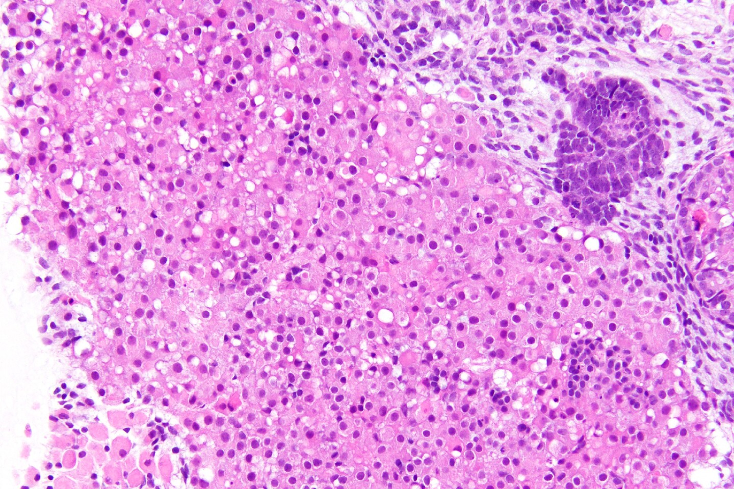
The structure and function of adrenal gland ‘organoids’ grown in a petri dish at the School of Veterinary Medicine closely replicated that of the human adrenal gland, according to a new study. (Image: Courtesy of the Sasaki laboratory)
Sitting atop the kidneys, the adrenal gland plays a pivotal role in maintaining a healthy body. Responding to signals from the brain, the gland secretes hormones that support critical functions like blood pressure, metabolism, and fertility.
People with adrenal gland disorders, such as primary adrenal insufficiency, in which the gland does not release sufficient hormones, can suffer fatigue, dangerously low blood pressure, coma, and even death if untreated. No cure for primary adrenal insufficiency exists, and the lifelong hormone-replacement therapy used to treat it carries significant side effects.
A preferable alternative would be a regenerative medicine approach, regrowing a functional adrenal gland capable of synthesizing hormones and appropriately releasing them in tune with the brain’s feedback. With a new study in the journal Developmental Cell, researchers from the University of PennsylvaniaSchool of Veterinary Medicine coaxed stem cells in a petri dish to divide, mature, and take on some of the functions of a human fetal adrenal gland, bringing that goal one step closer.
“This is a proof-of-principle that we can create a system grown in a dish that functions nearly identically to a human adrenal gland in the early stages of development,” says Kotaro Sasaki, senior author and an assistant professor at Penn Vet. “A platform like this could be used to better understand the genetics of adrenal insufficiency and even for drug screening to identify better therapies for people with these disorders.”
Postdoc Michinori Mayama examines a petri dish upon which adrenal organoids are growing. (Image: Courtesy of the Sasaki laboratory)
Sasaki says his team’s aim was to use human inducible pluripotent stem cells (iPSCs), which can give rise to a myriad of different cell types, to mimic the stages of normal human adrenal development. During this process, the cells would get directed to take on the characteristics of the adrenal gland.
To begin, the researchers used what’s known as an “organoid culture” system, in which cells grow first as a floating aggregate for three weeks, then on a membrane exposed to air on one side, promoting better survival and allowing them to proliferate in three dimensions. Utilizing a carefully selected growth medium, they prompted the iPSCs to elicit an intermediate tissue type in the adrenal development process, the posterior intermediate mesoderm (PIM).
After verifying they had cultured PIM-like cells, the researchers embarked on directing those cells to transition to the next stage, adrenocortical progenitor-like cells, during which cells turn on markers indicating they have “committed” to becoming adrenal gland cells.
Molecular assays to check for adrenal markers, as well as transmission electron microscope analyses, all told Sasaki and colleagues they were on the right track to recreating a tissue that resembled the early adrenal gland.
Kotaro Sasaki is a faculty member at the School of Veterinary Medicine.
“The process we developed was highly efficient, with around 50% of cells in organoids acquiring adrenocortical cell fate,” says Michinori Mayama, a postdoc in Sasaki’s lab and a lead author on the study. “The ovoid cells with voluminous pink cytoplasm and relatively small nuclei that we saw in our cultures are very characteristic of human adrenal cells at that stage.”
Sasaki, Mayama, and the rest of the research team performed a number of tests to evaluate how closely the functionality of the cells they had cultivated mirrored that of a human adrenal gland. They found the lab-grown cells produced steroid hormones, like DHEA, just as the “real-life” equivalent would. “In vitro, we can produce much of the same steroids that are produced in vivo,” Mayama says.
They also showed that the cells they grew could respond to what’s known as the hypothalamic-pituitary-adrenal axis, a feedback loop that governs communication from the brain to the adrenal gland and back again. “We used drugs that normally suppress adrenal DHEA production and showed that our iPSC-derived adrenal cells respond similarly to these drugs, with a marked reduction of hormone production,” says Sasaki. “This means that you can use this system for screening drugs that target adrenal hormone production, which could benefit patients with excessive adrenal hormone production or with a prostate cancer that exploits adrenal hormones for their growth.”
As the researchers refine their system, they say they hope to be able generate more of the gradations in tissue type that occur in a mature adult adrenal gland.
Adrenal organoids grown in the lab offer scientists the ability to ask new questions about the genetics behind adrenal gland disorders, test new drugs, and one day perhaps even offer cell therapies that could stand in for a malfunctioning adrenal gland. (Image: Courtesy of the Sasaki laboratory)
Such a platform opens up opportunities to learn far more about the still-mysterious adrenal gland. In particular, Sasaki notes that it could be leveraged to probe the genetic basis of adrenal insufficiencies as well as other diseases, such as adrenal carcinomas. Ultimately, the approach used to create this gland-in-a-dish may one day work to reconstitute a functioning brain-adrenal gland feedback loop in people with adrenal gland disorders.
“This is a first-of-its kind study,” says Sasaki. “The field of cell therapy holds so much promise for treating not just adrenal insufficiencies but other hormone-driven diseases: hypertension, Cushing syndrome, polycystic ovary syndrome, and more.”
Kotaro Sasaki is an assistant professor in the Department of Biomedical Sciences in the University of Pennsylvania School of Veterinary Medicine.
Michinori Mayama is a postdoctoral fellow in Penn Vet’s Department of Biomedical Sciences.
Sasaki and Mayama’s coauthors were Penn Vet’s Yuka Sakata, Keren Cheng, and Yasunari Seita; Penn Medicine’s Andrea Detlefsen, Clementina A. Mesaros, Trevor M. Penning, Wenli Yang, and Jerome F. Strauss III; Kyosuke Shishikura of Penn’s Department of Chemistry; and Richard J. Auchus of the University of Michigan. Sakata, Cheng, and Mayama were co-first authors on the study. Sasaki was senior author.
The study was supported, in part, by the Silicon Valley Community Foundation (Grant 2019-197906) and the Good Ventures Foundation (Grant 10080664).
Nanoparticle blueprints reveal path to smarter medicines
New research involving Penn Engineering shows detailed variation in lipid nanoparticle size, shape, and internal structure, and finds that such factors correlate with how well they deliver therapeutic cargo to a particular destination.
A generous gift from alumni Glenn and Amanda Fuhrman brings the work of internationally acclaimed artist Jaume Plensa to the University of Pennsylvania. The latest addition to the Penn Art Collection expands Philadelphia's public art.
A massive chunk of ice, a new laser, and new information on sea-level rise
For nearly a decade, Leigh Stearns and collaborators aimed a laser scanner system at Greenland’s Helheim Glacier. Their long-running survey reveals that Helheim’s massive calving events don’t behave the way scientists once thought, reframing how ice loss contributes to sea-level rise.


