How does opioid exposure affect brain development in young children?
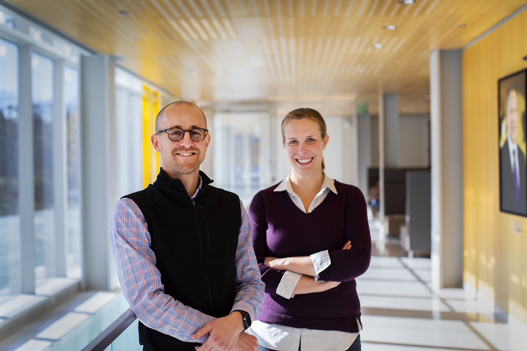
That’s the question Allyson Mackey and Dylan Tisdall hope to answer, through a new grant from an NIH initiative focused on addiction research.
The new NIH-funded work from researchers Dylan Tisdall of Penn Medicine and Allyson Mackey of the School of Arts and Sciences will work to develop an MRI method geared specifically to three- to five-year-olds and calculate how exposure to opioids can impede neurocognitive development of children in that age range.
As the opioid crisis has worsened in the past two decades, there’s been significant attention paid to how the drugs physically influence users. But what’s less understood is how opioid exposure affects the brain development of their children during infancy, toddlerhood, and beyond.
Partially, that’s because the best way to study what’s happening in the brain is through neuroimaging via an MRI. To produce clear, readable images, a person must lay extraordinarily still during scanning—a tall order for anyone, but especially for a restless five-year-old.
“It’s clear that the NIH wants to study greater adversity and kids who are younger,” says Tisdall, a researcher at the Perelman School of Medicine. “So, it’s essential that we improve and validate these tools.”
Most people understand intuitively that when you’re trying to take a picture and you move, the picture ends up blurry. It’s the same problem with an MRI.
Allyson Mackey, assistant professor in Penn Psychology
For nearly a decade, Tisdall has been thinking about how to make MRIs less sensitive to movement. He co-invented a motion-correction technology that can make in-the-moment adjustments when a subject moves; it’s currently being employed in two long-term, multicenter NIH projects, the Adolescent Brain Cognitive Development (ABCD) study and the Human Connectome Project. Mackey, a psychologist who studies how environmental factors affect brain plasticity in children, has been using neuroimaging in her work for years.
“It’s a really big problem in early-development neuroimaging when kids move. We get data that’s not very useable,” Mackey says. “Most people understand intuitively that when you’re trying to take a picture and you move, the picture ends up blurry. It’s the same problem with an MRI.”
“Except our exposure time is about six minutes,” Tisdall adds. “It’s like old-time photography.”
For this reason, the brain scan of a child this age traditionally happens during naptime or at night. Putting a sleeping child into the machine removes the movement challenge, producing clear pictures of the brain’s structure. But it doesn’t tell researchers anything about how the brain is functioning when that child concentrates on tasks or experiences emotions—key facets in understanding the impact of opioid exposure. For that, the child needs to be awake.
During the year and a half this project will run, Mackey and Tisdall plan to scan 100 awake and squirmy three- to five-year-olds. The feasibility study will compare the existing MRI methods used in the ABCD study with new ones the Penn researchers will develop that are geared toward this age range. Through partnerships with community organizations, they hope to recruit participants from the Kensington neighborhood in northeast Philadelphia, a place particularly hard-hit by the opioid crisis.
Each family’s single half-day visit will involve the researchers playing games with the child, including using a pretend MRI they refer to as the “elevator bed.” The child sits in the mock scanner watching a movie, which pauses every time there’s movement of any kind. It’s a simple way to reinforce the benefits of staying still in this context, Mackey says.
“They don’t actually know what most of this means, so we have them practice,” she says. “For the scan itself, we target 30 minutes, tops, so as short as possible.”
Though there hasn’t yet been extensive research done in this area, it’s clear there’s a link between exposure to opioids and a greater risk of attention deficit hyperactivity disorder, anxiety, low academic performance, and poor health. The Penn team also wants to look at how drug use affects the parent-child relationship. “We know there’s an increased risk of neglect,” Mackey says, “and there’s evidence to show that neglect does affect how the brain develops in young children.”
The funding Mackey and Tisdall received is part of more than $22 million from NIH HEAL that went to Penn researchers, who will study areas like pain management, prevention of opioid addiction, and novel medication options for opioid use disorder and overdose. It’s also Phase I of an NIH study called the HEALthy Brain and Child Development Study, which will follow 10,000 children from birth until age 10, including a large cohort exposed to opioids prenatally. Phase II is set to begin in 2021.
For now, Mackey and Tisdall are focused on Phase I because the data they collect could start to unravel some of the mysteries behind how children’s experiences and environment relate to their brain development. “We’re excited to be able to bring in 100 kids, to try out these new technologies and potentially get new data,” Tisdall says. “Hopefully, we can start to get some preliminary answers to the big scientific questions.”
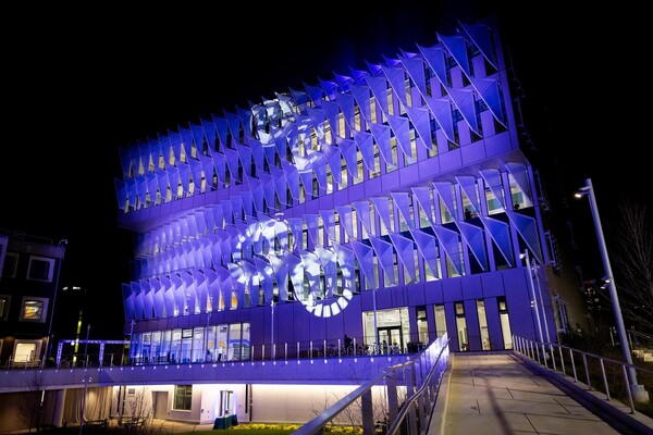
In Senegal, the ambitious Dakar Greenbelt project seeks to create an extensive network of ecological infrastructure in and around the city to sustainably address environmental concerns and enhance urban life. With support from David Gouverneur and Ellen Neises, Ph.D. candidate Rob Levinthal in the Weitzman School of Design led two courses that included a field trip to Dakar, that culminated in students presenting their visions for parts of the Greenbelt.
From a desert to an oasis: Penn engages in ambitious greening effort in the Sahel
Students from the Weitzman School of Design journeyed to Senegal to help with a massive ecological and infrastructural greening effort as part of their coursework. The Dakar Greenbelt aims to combat desertification and promote sustainable urban growth.
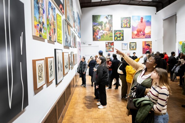
As part of an undergraduate course, Penn faculty and students curated an Arthur Ross Gallery exhibition of works from the Neumann family’s extensive collection of modern and contemporary art.
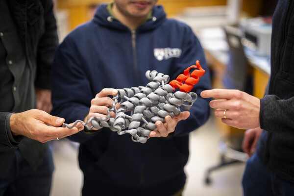
The University’s nexus for technology transfer supports researchers in their innovative efforts, from CAR T to mRNA advancements that have dramatically reshaped the world.